Showing posts with label Pathology. Show all posts

Wednesday, 2 September 2020

Pathology : High Yield

 1.  Damage  to  nervous  tissue  is  repaired  by  :  Neuroglia

 2.  Mycosis  fungoides  :  Cutaneous  lymphoma   

3.  Secondary  amyloidosis  complicates:  Chronic  osteomyelitis 

4.  The  earliest  feature  of  TB  is:  Lymphocytosis   

5.  The  low  grade  non-hodgkins  lymphoma  is  :  Follicular  

 6.  Liquefactive  necrosis  is  seen  in  :  Brain  

 7.  The  crescent  forming  glomerulonephritis  is:  RPGN 

8.  Earliest  feature  of  correction  of  IDA  is  :  Reticulocytosis

   9.  Kupffer's  cells  are  found  in  :  Liver   

10.  Heart  failure  cells  are  found  in  :  Lungs 

  11.  Psammoma bodies  show:  Dystrophic  calcification   

12.  Beta-microglobulin:  is  not  a  tumor  marker  

 13.  Commonest  benign  tumor  of  liver  :  Hemangioma  

 14.  Blood  when stored  at  4  degree  celcius  can  be  kept  for:  21  days   

15.  Congo-red  with  amyloid  produces:  Brilliant  pink  ccolour

  16.  Cloudy  swelling  does  not  occurs  in  :  Lungs 

17.  Gamma Gandy bodies contains  hemosiderin  and  :  Ca++   

18.  Hutchinson's  secondaries  in  skull  are  due  to  tumors  in  :  Adrenals   

19.  Albumino  -cytologic  dissociation  occurs  in  cases  of:  Guillain  Barre  syndrome   

20.  Metastatic  calcification  is  most  often  seen  in  :  Lungs   

21.  ASLO  Titres  are  used  in  the  diagnosis  of:  Acute  rheumatic  fever  

 22.  Apoptosis  is  inhibited  by  :  bcl-2  

 23.  CEA:  is  not  used  as  a  tumor  marker  in  testicular  tumours

 24.  Onion  peel  appearance  of  splenic  capsule  is  seen  in  :  SLE
Monday, 18 November 2019

DOC Cancers

* DOC for CLL-- Fludarabin
* DOC for Brain Tumors- Temozolamide
* DOC for Pancreatic Carcinoma-- Gemcitabine
* DOC for Choriocarcinoma- Methotrexate
* DOC for Colorectal cancer-- 5-FU
*DOC for Hairy Cell Leukemia- Cladribine
*Drugs of choice for Wenger's granulomatosis-- Cyclophosphamide
*DOC for Multiple Myloma-- Bortezomib

Tuesday, 12 June 2018

Tetralogy of Fallot, demystified


Tetralogy of Fallot is a fairly common heart defect. In fact, it's the most common cyanotic heart defect (meaning that if a baby is born with cyanosis - markedly decreased oxygen saturation - and the cause is determined to be a congenital heart defect, the most likely culprit is tetralogy of Fallot).

From the name, it’s obviously composed of four parts. But how to remember what those parts are? You could just memorize them using brute force, but there's actually one thing that ties them all together - so if you can remember this one thing, then the four things make sense. I love this, because when you find you've forgotten one of the four things (which you probably will), you can actually reason it out. Hurray!

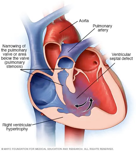
Here's the one thing to remember, and it's actually the thing that causes the whole disease. When the interventricular septum is forming, the top portion is pushed up and towards the right ventricle. The official name for this is anterosuperior displacement of the infundibular septum, but it's easier to just remember "up and towards the right," I think.


Check out this image of tetralogy of Fallot. See the red asterisk? That's the top of the interventricular septum. The black asterisk marks the bottom of the interventricular septum. Those parts are supposed to be connected...but obviously they're not. And the reason they're not is that the top part of the septum has moved up (creating a hole in the septum) and to the right (smushing the pulmonary artery outflow tract).

So what happens as a result?

One of the main problems is that it's now very hard for the blood to get out of the right ventricle and into the pulmonary artery (and lungs). That's because the pulmonary outflow tract (fancy name for the beginning of the pulmonary artery) is compressed, and there's less room for blood to flow through. This is called pulmonary stenosis. This is what causes the baby to be cyanotic! If you can't get enough blood out into the lungs, the blood isn't going to be oxygenated very well, and the baby's skin will be bluish. The right ventricle has to work hard to push blood through that compressed pulmonary artery - so the right heart becomes hypertrophied (bigger).

A couple other things happen too, as a result of this displacement of the septum. As the top part of the septum moves upwards, it separates from the bottom part, creating a hole in the septum (this is called a ventricular septal defect). This is actually kind of a good thing, in this case, because it relieves a little of the pressure on the right side of the heart. If the septum was intact, then the only place for the right ventricular blood to go would be through the compressed pulmonary artery...and the right ventricle would have to work incredibly hard to empty itself with each cardiac cycle.

Finally, as the top part of the septum moves to the right, it pulls the aortic valve along with it, repositioning it so that it sits pretty much right over the ventricular septal defect. This is called an overriding aorta, and it doesn't have much clinical consequence.  

So to summarize: the top of the septum moves up and to the right, causing:
1. Pulmonary stenosis 
2. Right ventricular hypertrophy 
3. A ventricular septal defect
4. An over-riding aorta  

Tuesday, 29 August 2017

Forebrain developmental abnormalities

Megalencephaly
  • Abnormally large brain
  • Less common than microencephaly 
Microencephaly
  • Abnormally small brain
  • Lots of causes (chromosomal abnormalities, fetal alcohol syndrome, HIV acquired in utero)
Lissencephaly
  • Decrease in number of gyri
  • Sometimes gyri are totally absent (agyria)
Polymicrogyria
  • Small, numerous, irregularly formed gyri
  • Can be caused by injury or genetic abnormality
Neuronal heterotopias
  • Neurons in inappropriate locations along migrational pathways
  • Associated with epilepsy
Holoprosencephaly
  • Incomplete separation of cerebral hemispheres across the midline
  • May have midline facial abnormalities (e.g., cyclopia, absence of olfactory cranial nerves)
Agenesis of the corpus callosum

  • Absence of white matter bundles connecting cerebral hemispheres
  • Patients may have other defects, or may be asymptomatic

Sunday, 19 February 2017

Microarray

MICROARRAY Analysis:
__________________________
RNA is prepared from cells, reverse transcribed to cDNA, and labelled with fluorescent dyes .
Green dye for normal and red dye is for cancer cells.
The fluorescent probes are mixed and hybridized to a cDNA array.
Each spot on the array is an oligonucleotide (or cDNA fragment) that represents a different gene.
Image is captured by fluorescence camera where:
a) Red spot indicates higher expression in tumor cells.
b) Green spot indicates lower expression in tumor cells.
c) Yellow spot indicates equal expression level in both normal and tumor samples

Flow cytometry markers :

Flow cytometry markers :
CD1a, S-100, CD-207(Langherin)- Langerhan cell histiocytosis
CD 2 3 4 5 7 8- T-cell markers,CD3 is pan T cell marker
CD10 (aka CALLA antigen) : Early pre B marker,immature B cell marker
CD 11c, 25, 103, 123 : Hairy cell lukemia (CD 123 Most specific)
CD 13,33,117, MPO: Myeloid series marker-AML
CD 14,64: Monocyte marker (AML-M4 M5)
CD 15 : RS cell,Neutrophils
CD 15,30 : RS Cell
CD 16,56: NK Cell
CD 19,20,21,22 : B Cell markers, CD 19 is pan B cell marker
CD 23+,CD 5+ : CLL/SLL
CD 23-,CD 5+ : Mantle cell lymphoma
CD 30(only) aka Ki-antigen : Anaplastic Large cell lymphoma
CD 31: Endothelial cell marker (positive in angiosarcomas)
CD 34: Stem cell (also positive in angiosarcomas)
CD 41,61: Megakaryocyte,platelet marker,positive in AML-M7
CD 45 : On all leukocytes(except RS cell)
CD 45 RO: Memory cell
CD 45RA/RB: Naive B/T Cell
CD 55(DAF), CD 59(MIRL) : Absent in PNH
CD 68,S-100+ :Histiocyte marker(+ in malignant fibrous histiocytosis)
CD 95/FAS ligand: apoptosis marker (extrinsic pathway)
CD 99/MIC-2 : Ewings sarcoma
CD 103: Hairy cell
CD 117 :GIST,Mastocytosis,AML (117 is present on mast cells also)
CD 123: New antibody in Hairy cell leukemia
CD 133:Glioma
CD 207(Langherin): LCH
CD 235(Glycophorin): AML-M6

Saturday, 5 November 2016

Important stains and their uses in pathology.

Important stains and their uses in pathology.
1. Lipid or fat - Sudan black or oil red O
2. Calcium - von kossa or alzarine red s
3. Haemosiderin or iron - Prussian blue or Perls stain
4. Collagen - Masson trichrome
5. Mucin - mucicarmine, alcian blue
6. Fungi - PAS, silver methanamine
7. Melanin - Masson Fontana
8. Mast cells - toluidine blue
9. Amyloid - Congo red
10. Basement membrane - PAS
11. Nucleic acid or DNA - feulgen, acridine orange
12. Routine histo pathology - hematoxylin and eosin ( H&E)
13. Routine peripheral smear - geimsa or leishman
14. Lymphoblast - PAS ( block positivity)
15. Myeloblasts - myeloperoxidase, non specific esterase, Sudan black B.

Saturday, 26 December 2015

AMBIGUOUS LEUKEMIA

AMBIGUOUS LEUKEMIA

3 TYPES:

1) UNDIFFERENTIATED ACUTE LEUKEMIAS:
Lack morphologic or immunologic differentiating features;
Blasts cell express HLA-DR,CD34,CD38,TdT( terminal deoxynucleotidase) and CD7.

2) BILINEAL ACUTE LEUKEMIA:
Two populations of blasts expressing distinct lineage markers of myeloid or lymphoid origin or B- and T-cell lineage

3) BIPHENOTYPIC ACUTE LEUKEMIA:
Same Blast cell coexpress myeloid and lymphoid antigen both ..usually seen in BLAST CRISES OF CHRONIC MYELOID LEUKEMIA

Wednesday, 23 December 2015

HLA

HLA diseases are mainly due to HLA A , HLA B, HLA-DR

HLA B-27

very popular mnemonic : PAIR from first Aid

P soriasis

A nkylosing spondylitis
I nflammatory bowel disease
R eiter’s syndrome.
………………………………………

HLA B-8 : Graves disease
In GRAVE yard maggots BITE (B8), So Grave is B8

………………………………………………….
HLA-DR2 :

A person looks into his mobile and then tells to his wife : Hey MSG about Appointment to DR

HeY – Hay fever
M – Multiple Sclerosis
S – SLE
G – Goodpasture’s
……………………………
HLA-DR4 : Rheumatoid Arthritis

Rheumatoid Arthritis Affects Joints and We have Four large Joints in our body like two shoulder and two hip Joints.
……………………………………….
HLA- DR5:

Hush Puppies : very popular footwear

Hush : Hasimoto’s Thyroiditis

Puppies : Pernicious Anemia ( P for Pentad = 5 )
…………………………………………………
HLA -DR7

My 7th Sense says that Nephrotic Syndrome responds to Steroid

Steroid responsive Nephrotic Syndrome
………………………………
HLA- DR3-DR4

3 -4 I want More ( Diabetic patient Eats more )

Type 1 DM
………………………..
HLA -A3

Hemochromatosis

Iron has 3 positive charges (+3) and “He ” Sounds like “Ae” So A3 in Hemochromatosis .

Saturday, 12 December 2015

Prothrombin

Prothrombin.
☆Intrinsic and Extrinsic pathways of coagulation converge at factor 10.
☆Threonine does not contain Sulfa group.
☆Autosomal dominant is hereditary Spherocytosis & Poly cystic kidney disease.
☆Lens opacity causing drugs >Chlorpromazine, Amidarone, Tamoxifen, Gold & Iron toxicity.
☆Drugs causing corneal opacity > Amiodarone, Chloroquine, Mepacrine & Copper.
☆ Ribosome have purple color on Eosin & Methylene blue staining
☆High energy content > Starch.
☆High energy compound > ATP
☆Antidote of warfarin is vitamin K but if action is more quickly required then FFP.
☆Olfactory cells are the only neurons in the body that regenerates.
☆Projectile vomiting greenish in color means bilious vomiting so it is due to duodenal atresia but if projectile vomiting non bilious then it is hypertrophic pyloric stenosis.
☆ Pulmonary trunk relation with the bronchus at the hilum of the lung-mnemonic is RALSR- Right Anterior & Left Superior.
☆Rhino sinusitis is caused by Strep Pneumonia, H. Influenza, M Catarrahalis.
☆ Homan’s sign is present in DVT in which if you dorsiflex the foot there will be pain in calf muscles.
☆ Classic triad of Pulmonary Embolism: -
☆☆Neurological manifestations.
-Petechial rash.
-Hypoxemia.
☆Nitrogen bubbled precipitator in ascending divers and can be treated with hyperbaric oxygen.
☆CT pulmonary angiography is the best test to detect Pulmonary Embolism.
☆The most common infectious agent transmitted by blood transfusion is cytomegalovirus (CMV), which is present in donor lymphocytes.
☆Before blood is transfused into newborns or patients with T-cell deficiencies, it must be irradiated to kill donor lymphocytes. This prevents the patient from developing a graft-versus-host reaction or a CMV infection.
☆Yersinia enterocolitica, a pathogen that thrives on iron, is the most common contaminant of stored blood.
☆ Iron is stored in bone macrophages.
☆ Structures passing thru superior orbital fissure....
NOT-FAL
¤NASOCILLIARY¤OPTHALIMIC VEIN¤TROCHLEAR¤FRONTAL
☆☆Suture Removal:
¤Head 5-7days
¤Face 3-5days
¤Eyelid & eyebrow 3-5days
¤Trunk 5-7days
¤Extremities 7-10days
¤Surface of joint 10-14days
¤Hand ==7days
☆☆Absorption
☆iron nd divalents absorb in duodenum.
☆Folic acid, maximum water, max electrolytes, long chain fatty acids in jejunum.
☆Bile nd B12 absorb in ileum.
☆water nd electrolytes absorb in colon but less than jejunum. Short chain fatty acids absorb in colon.
¤¤Buffers
☆Major intracellular buffer is protein.
☆Major extracellular buffer is bicarb.
☆If only major buffer asked then Bicarb.
☆Major renal buffer is still bicarbonate if depleted theb
¤Phosphate ==Qualitative
¤Ammonia == Quantitative
☆Buffer in blood is H2co3 > Hb
¤¤Uterus
☆Uterosacral felt on PR
☆Main support is cardinal(also named transverse cervical ligament )
☆Round ligament of uterus keeps it anteverted anteflexed
☆broad ligament has very lessor role in support
☆Best way to "measure" gfr is inulin clearance.
☆best way to "estimate" gfr is creatinine clearance.
☆best way to "clincally" measure gfr is creatinine clearance.
☆best way to measure renal plasma / blood flow is PAH .
☆best test for renal falilure is creatinine clearance.
☆☆☆☆ blood transfusion
↪multiple===hemochromatosis
↪massive===hyperkalemia
↪repeated ===hypocalcemia.

Saturday, 21 November 2015

Blood Transfusion

THE RIGHT WAY OF ADMINISTERING BLOOD PRODUCTS

[ from "THE CLINICAL USE OF BLOOD: HAND BOOK , World Health Organization & Blood Transfusion Safety , GENEVA ]

✔️Prefer a larger cannula: A doubling of the diameter of the cannula increases the flow rate of most fluids by a factor of 16.

✔️In case of Whole blood, red cells, plasma and cryoprecipitate
>Use a new, sterile blood administration set containing an integral 170–200 micron filter

>Change the set at least 12-hourly during blood component infusion
>In a very warm climate, change the set more frequently and usually after every four units of blood, if given within a 12-hour period

✔In case of Platelet concentrates

>Use a fresh blood administration set or platelet transfusion set, primed with saline.

✔️WARMING BLOOD:

>There is no evidence that warming blood is beneficial to the patient when infusion is slow.

>At infusion rates greater than 100 ml/minute, cold blood may be a contributing factor in cardiac arrest. However, keeping the patient warm is probably more important than warming the infused blood.

>Warmed blood is most commonly required in: [1]Large volume rapid transfusions:
    -Adults: greater than 50 ml/kg/hour      -Children: greater than 15 ml/kg/hour
[2]Exchange transfusion in infants  [3]Patients with clinically significant cold agglutinins.

>Blood SHOULD ONLY BE WARMED in a blood warmer. Blood warmers should have a visible thermometer and an audible warning alarm and should be properly maintained.

>Blood should never be warmed in a bowl of hot water as this could lead to haemolysis of the red cells which could be life-threatening.

✔️Severe reactions most commonly present during the first 15 minutes of a transfusion. All patients and, in particular, unconscious patients should be monitored during this period and for the first 15 minutes of each subsequent unit.

✔️The transfusion of each unit of the blood or blood component should be completed within four hours of the pack being punctured. If a unit is not completed within four hours, discontinue its use and dispose of the remainder through the clinical waste system.

Monday, 21 September 2015

Abscopal Effect

Splenectomy is an effective secondary or tertiary treatment for two chronic B cell leukemias, hairy cell leukemia and prolymphocytic leukemia, and for the very rare splenic mantle cell or marginal zone lymphoma.

Splenectomy in these diseases may be associated with significant tumor regression in bone marrow and other sites of disease.

Similar regressions of systemic disease have been noted after splenic irradiation in some types of lymphoid tumors, especially chronic lymphocytic leukemia and prolymphocytic leukemia.

This has been termed the abscopal effect. Such systemic tumor responses to local therapy directed at the spleen suggest that some hormone or growth factor produced by the spleen may affect tumor cell proliferation.

Friday, 11 September 2015

Myelodisplastic Syndrome

MYELODYSPLATIC SYNDROME Asked Points :
MC cytogenetic change in Myelodysplastic Syndrome in adult - 5q deletion
Cytogenetic change in Myelodysplastic Syndrome in children - Monosomy 7
Common age group affected by Myelodysplastic syndrome - >50
Ringed sideroblast characteristically seen in - Myelodysplastic syndrome
Pawn ball megakaryocytes is characteristic of - Myelodysplastic syndrome
NOT true about Myelodysplastic syndrome - Hypocellular bone marrow
Act by hypomethylation - Decitabine
Harrison Points :
Megaloblastoids are seen in - Myelodysplasia

Ringed sideroblast - Mitochondria encrusted iron
International prognostic scoring system for - Myelodysplasia
Drug used for myelodysplastic syndrome - Lenalidomide
Additional Points :
Treatment of MDS with 5q deletion - Lenalidomide

Monday, 7 September 2015

TRALI

TRALI ( Transfusion related acute lung injury )
1. Definition - it is acute lung injury which occurs within 6 hours of completion of transfusion of blood components.
Pathogenesis - two hit hypothesis.
First hit is the priming event like endothelial activation which leads to increased sequestration and
sensitisation of neutrophils in the micro vasculature of lung. Second hit can be the antibodies in
the transfused blood product that recognise antigens expressed on neutrophils. Most common
antibodies can be those that bind MHC antigens.
2. TRALI can occur with all plasma containing products but more common with transfusion of FFP and platelets.
3. Clinically - dyspnea, tachypnea, fever, hypotension.
4. Chest X Ray - bilateral pulmonary infiltrates or white out lung. That is why most important differential diagnosis is ARDS.

Friday, 4 September 2015

Myelodysplastic Syndrome

MYELODYSPLATIC SYNDROME
Asked Points :
MC cytogenetic change in Myelodysplastic Syndrome in adult - 5q deletion
Cytogenetic change in Myelodysplastic Syndrome in children - Monosomy 7
Common age group affected by Myelodysplastic syndrome - >50
Ringed sideroblast characteristically seen in - Myelodysplastic syndrome
Pawn ball megakaryocytes is characteristic of - Myelodysplastic syndrome
NOT true about Myelodysplastic syndrome - Hypocellular bone marrow
Act by hypomethylation - Decitabine
Harrison Points :
Megaloblastoids are seen in - Myelodysplasia
Ringed sideroblast - Mitochondria encrusted iron
International prognostic scoring system for -
Myelodysplasia
Drug used for myelodysplastic syndrome -
Lenalidomide
Additional Points :
Treatment of MDS with 5q deletion - Lenalidomide

Coombs Test

A) DIRECT COOMBS TEST:-

(also known as the direct antiglobulin test or DAT)

● detect antibodies or complement bound to RBC surface antigens in vivo.

●used for:-
1) immune-mediated hemolytic anemia
2) Hemolytic disease of the newborn
3) Rh D hemolytic disease of the newborn
4) ABO hemolytic disease of the newborn
5) Drug-induced immune-mediated hemolysis
6) Transfusion reaction, such as one due to improperly matched units of blood

B) INDIRECT COOMBS TEST :-

(also known as the indirect antiglobulin test or IAT)
●detect in-vitro antibody-antigen reactions

●used for:-

1) detection of very low concentrations of antibodies present in a patient's plasma/serum prior to a blood transfusion
2) in antenatal care, the IAT is used to screen pregnant women for antibodies that may cause hemolytic disease of the newborn
3) compatibility testing
4) antibody identification
5) RBC phenotyping
6) titration studies.


?☺: Tumor Lysis Syndrome

* Most commonly seen with acute leukemias, Burkitt lymphoma and other lympho-reticular malignancies (uncommon in solid tumors).
* Chemotherapy results in death of large quantities of cells, which leads to release of massive amounts of potassium, uric acid and other breakdown products into the blood.
* It develops within hours to few days of initiation of chemotherapy.

* Features:
- hypocalcemia
- renal failure due to precipitation of uric acid crystals or calcium phosphate crystals in the kidney
- hyperkalemia leading to arrhythmias
- hyperphosphatemia

* Management
- vigorous IV hydration with half normal saline
- correction of metabolic abnormalities
- hemodialysis in severe cases

Friday, 14 August 2015

Gauchers Disease

Gaucher's disease-
M/C lysosomal storage disease* with *beta-glucosidase deficiency.

Types-• Non-neuropathic: Type 1 (M/C)• Neuropathic: Type 2,3 (Incompatible with life)

C/F:• Hematology-
◦ Anemia◦ Thrombocytopenia
• Organomegaly• Bony lesion

Histopathology-◦ Gaucher'scell-▪ Blendnucleus▪ Wrinkledpaperlikecytoplasm ▪ PAS+ve▪ Oil-Red-Onegative

Enzyme replacement therapy available

CD Markers

All CD at one place

CD1a, S-100, CD-207(Langherin)-Langerhan cell
histiocytosis
CD 2 3 4 5 7 8- T-cell markers,CD3 is pan T cell marker
CD10 (aka CALLA antigen) : Early pre B marker,immature
B cell marker
CD 11c, 25, 103, 123 : Hairy cell lukemia (CD 123 Most
specific)
CD 13,33,117, MPO: Myeloid series marker-AML
CD 14,64: Monocyte marker (AML-M4 M5)
CD 15 : RS cell,Neutrophils
CD 15,30 : RS Cell
CD 16,56: NK Cell
CD 19,20,21,22 : B Cell markers, CD 19 is pan B cell
marker
CD 23+,CD 5+ : CLL/SLL
CD 23-,CD 5+ : Mantle cell lymphoma
CD 30(only) aka Ki-antigen : Anaplastic Large cell
lymphoma
CD 31: Endothelial cell marker (positive in angiosarcomas)
CD 34: Stem cell (also positive in angiosarcomas)
CD 41,61: Megakaryocyte,platelet marker,positive in AML-
M7
CD 45 : On all leukocytes(except RS cell)
CD 45 RO: Memory cell
CD 45RA/RB: Naive B/T Cell
CD 55(DAF), CD 59(MIRL) : Absent in PNH
CD 68,S-100+ :Histiocyte marker(+ in malignant fibrous
histiocytosis)
CD 95/FAS ligand: apoptosis marker(extrinsic pathway)
CD 99/MIC-2 : Ewings sarcoma
CD 103: Hairy cell
CD 117 :GIST,Mastocytosis,AML (117 is present on mast
cells also)
CD 123: New antibody in Hairy cell leukemia
CD 133:Glioma
CD 207(Langherin): LCH
CD 235(Glycophorin): AML-M6

Tumour Grading

Tumor grading
1. Bloom-Richardson grading- Breast Ca.
   T- Tubular formation
   N- Nuclear pleomorphism
   M- Mitotic count
2. Nottingham prognosis index- Breast
   (0.2*Tumor size+LN+Grade)
3. Chang- Medulloastoma
4. Noguchi- Adeno Ca. of Lung
5. Masoka- Thymoma
6. Shimida index- Neuroblastoma
7. Gleason- Prostate
8. Robson- Renal Cell Ca.
9. Jackson- Penile Ca.
10. Dukes- Colorectal
11. Nevin- Ca. Gall bladder

Thursday, 30 July 2015

ML & DOWN SYNDROME

ML & DOWN SYNDROME
1. Down Syndrome kids have 150 fold RISK of AML by 5 years
2. MC AML subtype in Down syndrome is AML M7.
3. AML M7 is the MC leukemia in Kids < 3 years with Down syndrome
4. Children with Down syndrome and AML comprise 10% of all pediatric AML cases.
5 . ALL INFANTS WITH DOWN SYNDROME ARE CONSIDERED HIGH RISK FOR AML.
6. HIGHEST RISK of AML in Down syndrome is between 1-3 years age.. Those kids >4years and getting AML have a SIGNIFICANTLY LESSER SURVIVAL RATE compared to those <4years of age
7. Mutation in GATA-1 gene is present in all Down syndrome kids with Transient Myeloproliferative Disorder (TMD) and AML M7.
8. IN GENERAL Down syndrome kids with AML have better prognosis than NON-Down syndrome KIDS with AML!!