Tetralogy of Fallot is a fairly common heart defect. In fact, it's the most common cyanotic heart defect (meaning that if a baby is born with cyanosis - markedly decreased oxygen saturation - and the cause is determined to be a congenital heart defect, the most likely culprit is tetralogy of Fallot).
From the name, it’s obviously composed of four parts. But how to remember what those parts are? You could just memorize them using brute force, but there's actually one thing that ties them all together - so if you can remember this one thing, then the four things make sense. I love this, because when you find you've forgotten one of the four things (which you probably will), you can actually reason it out. Hurray!
Here's the one thing to remember, and it's actually the thing that causes the whole disease. When the interventricular septum is forming, the top portion is pushed up and towards the right ventricle. The official name for this is anterosuperior displacement of the infundibular septum, but it's easier to just remember "up and towards the right," I think.
Check out this image of tetralogy of Fallot. See the red asterisk? That's the top of the interventricular septum. The black asterisk marks the bottom of the interventricular septum. Those parts are supposed to be connected...but obviously they're not. And the reason they're not is that the top part of the septum has moved up (creating a hole in the septum) and to the right (smushing the pulmonary artery outflow tract).
So what happens as a result?
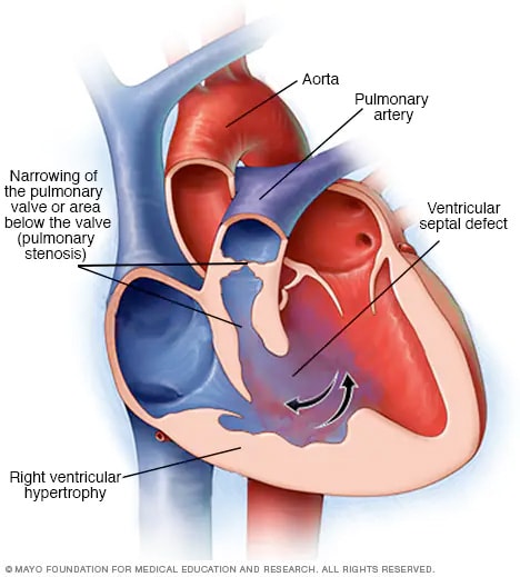
One of the main problems is that it's now very hard for the blood to get out of the right ventricle and into the pulmonary artery (and lungs). That's because the pulmonary outflow tract (fancy name for the beginning of the pulmonary artery) is compressed, and there's less room for blood to flow through. This is called pulmonary stenosis. This is what causes the baby to be cyanotic! If you can't get enough blood out into the lungs, the blood isn't going to be oxygenated very well, and the baby's skin will be bluish. The right ventricle has to work hard to push blood through that compressed pulmonary artery - so the right heart becomes hypertrophied (bigger).
A couple other things happen too, as a result of this displacement of the septum. As the top part of the septum moves upwards, it separates from the bottom part, creating a hole in the septum (this is called a ventricular septal defect). This is actually kind of a good thing, in this case, because it relieves a little of the pressure on the right side of the heart. If the septum was intact, then the only place for the right ventricular blood to go would be through the compressed pulmonary artery...and the right ventricle would have to work incredibly hard to empty itself with each cardiac cycle.
Finally, as the top part of the septum moves to the right, it pulls the aortic valve along with it, repositioning it so that it sits pretty much right over the ventricular septal defect. This is called an overriding aorta, and it doesn't have much clinical consequence.
So to summarize: the top of the septum moves up and to the right, causing:
1. Pulmonary stenosis
2. Right ventricular hypertrophy
3. A ventricular septal defect
4. An over-riding aorta
No comments:
Post a Comment