Showing posts with label General medicine. Show all posts

Monday, 18 November 2019

Respiratory System One liners

Respi system oneliners

[1] Creola bodies
In Bronchial asthma, air way epithelium is sloughed into bronchial lumen in form of CREOLA BODIES

[2] Asteroid body or Schaumann body
Nonspecific birefringent crystalline bodies in sarcoid granulomas.

[3] PULMONARY HEMORRHAGE primary causes:
[A] Good pasture syndrome
[2] Microscopic polyangitis
[C] Idiopathic pulmonary hemosiderosis

[4] Hypersensitivity pneumonitis : Important examples :-
[A] Bagassosis =  due to T actinomycetes from moldy bagasse (sugarcane)
[B] Bird fancier,s , breeder's , handler's lung = bird proteins from avian droppings.
[C] Farmers lung = T actinomycetes from moldy hey
[D] Malt workers lung = Aspergillus fumigatus from moldy barley

[5] Hypersensitivity pneumonits = Both type III and type IV hypersensivity reaction

[6] Caplan syndrome = Seropositive rheumatoid arthiritis + Progressive massive fibrosis
Initially it was described in coal workers but subsequebntly found in variety of pneumoconioses

[7] Byssinosis = (typical description to identify q) = is charecterised clinically by occasional in early stage and then regular (late stage) chest tightness toward the end of the first day of workweek (" MONDAY CHEST TIGHTNESS")

[8] Asbestosis usually affects lower lobe first then spreads upwards as disease becomes more severe whereas silocosis coal workers pneumoconiosis usually affects upper lobe.

[9] Presence of hilar lymphedenopathy or calcified hilar lymphnode " egg shell calcification" in a pneumoconioses is more suggestive of SILICOSIS among all other pneumoconiosis

[10] The single most useful clinical sign of the severity of pneumonia is ?
Ans = respiratory rate >30 /min [ref : hson]
Other scoring methods for severity of pneumonia= [a] CURB score [b] PORT risk class

[11] Guessing most likely etiological agent of pneumonia by radiological finding
[A] upper lobe cavity = tb
[B] pneumatocoele = s pneumoniae
[C] air fluid level = abscess = polymicrobial
[D] ***  CRESCENT SIGN in lung = Aspergillosis

[12] Pneumonia + hyponatremia = legionella pneumonia
[13] Hecht's pneumonia= interstitial giant cell pneumonia = measeles

[14] Triads 
SAMTER TRIAD = asthma + allergy + nasal polyposis
KARTARGENERS SYNDROME = situs inversus + bronchiectasis + sinusitis

[15] Friedlanders pneumonia = kleibsiella pneumonia = bulging fissure sign

Monday, 25 March 2019

Rule of 2 - Tuberculosis

Rule of two

1.      2 out of 10 word cases of TB are in India
2.      Tuberculosis has 2 words T and B = TB
3.      TB can be pulmonary or extra pulmonary.
4.      Resistant TB can be MDR or XDR Tb.
5.      MDR TB is seen in 2% of new TB cases.
6.      2 patients of every 10 patients are extra pulmonary RB cases
7.      One can treat both sputum positive of sputum negative cases
8.      Sputum negative cases confirm by GeneXpert or LPA test

9.      Don’t ignore fever and cough.

10.  Don’t ignore fever of more than 2 weeks duration.

11.  Don’t ignore cough of more than 2 weeks duration.

12.  Get 2 sputum examination to rule out TB (stat and nearly morning).


13.  Collect 2 ML of sputum for examination.


14.  Sputum can be tested by Z N Stain or fluorescent microscopy.

15.  2 tests for TB to remember: Sputum AFB and Molecular tests.


16.  Molecular tests can be GeneXpert TB test or LPA (line probe assay) test.


17.  GeneXpert TB tests is for rifampicin resistance (2R)


18.  GeneXpert TB test in private sector costs 2 thousand rupees.


19.  LPA for 2 drug resistances: INH and Rifampicin


20.  GeneXpert TB test results are available in 2 hours

21.  LPA test takes more than 2 days for the results


22.  In retreatment cases do both sputum AFB and molecular test


23.  In retreatment cases before labelling as a resistant case do the GeneXpert test 2 times.


24.  2 advanced investigations to remember: HRCT Chest and Bronchoscopy

25.  MDR TB means resistance to Rifampicin and INH. (2 drugs)


26.  XDR Tb means resistance to 2+2 drugs (Rifampicin and INH + quinolone and one injectable) or to 2 group of drugs


27.  Take 2 weeks precautions if AFB is positive.

28.  Rule out HIV and Diabetes Mellitus in case of TB.

29.  Use 2 type of masks, N 95 (for doctor) and surgical mask (for patients).

30.  Notify TB and screen the contacts.

31.  Not notifying TB is a crime under 2 clauses of MCI (5.2 and 7.14).

32.  Municipal Corporation and West Bengal CEA can take action if one fails to notify TB or fails to screen the contacts.

33.  Spreading infection is punishable under IPC Sections IPC 269/270.


34.  Patients of TB can be new cases or retreatment cases.


35.  There are 2 spells of treatment Intensive phase and continuation phase.


36.  For treatment TB can be sputum conformed TB or clinical TB.

37.  Give four drugs for 2 months.


38.  When we give 5 drugs we add SM injection for 2 months.

39.  At 2 months get 2 sputum tests done to confirm if AFB is negative or not.


40.  At 2 months if sputum AFB is positive think of GeneXpert TB tests or LPA test.


41.   In health care setting masks should be available at the reception and laboratory.

42.  Avoid split ACs at 2 places - in cars and at home.

43.  Use burial method or phenol to destroy the sputum.

44.  Skin and GI are the major organs affected by ATT.

45.  Vit B 6 and Vit D should be added to ATT.

46.  Brain and bone TB involvement require long treatment.


47.  MDR TB requires 2 years treatment.


48.  In MDR TB 2 out of 10 cases will die.


49.  In MDR cases 2 out of 10 will default.

50.  Patient rights: To get free diagnostics and free drugs from the government.


51.  Acid-fast bacteria visualized on a slide may represent M. tuberculosis or nontuberculous mycobacteria (NTM).


52.  Notify both confirmed and suspected case.


53.  Collect sputum: Spontaneously (by coughing) or induced by inhalation of aerosolized hypertonic saline generated by a nebulizer.


54.  Two most common presentation of Extra pulmonary TB are TB Lymph Nodes or pleural effusion.


55.  Common test for pleural effusion are proteins and presence of lymphocytes.


56.  Tow thigs to remember in pleural effusion: GeneXpert is negative and ADA test is positive.


57.  Paradoxical reaction in TB lymph nodes occurs at 2 months.


58.   MDR TB can be both pulmonary and extra pulmonary TB.


59.  Think before you order two tests: ELIESA and Gold Interferon.


60.  All biopsy samples should be taken in saline (for IRL) and formalin.


(with inputs from Dr Ashwani Khanna)

Tuesday, 12 June 2018

Tetralogy of Fallot, demystified


Tetralogy of Fallot is a fairly common heart defect. In fact, it's the most common cyanotic heart defect (meaning that if a baby is born with cyanosis - markedly decreased oxygen saturation - and the cause is determined to be a congenital heart defect, the most likely culprit is tetralogy of Fallot).

From the name, it’s obviously composed of four parts. But how to remember what those parts are? You could just memorize them using brute force, but there's actually one thing that ties them all together - so if you can remember this one thing, then the four things make sense. I love this, because when you find you've forgotten one of the four things (which you probably will), you can actually reason it out. Hurray!

Here's the one thing to remember, and it's actually the thing that causes the whole disease. When the interventricular septum is forming, the top portion is pushed up and towards the right ventricle. The official name for this is anterosuperior displacement of the infundibular septum, but it's easier to just remember "up and towards the right," I think.


Check out this image of tetralogy of Fallot. See the red asterisk? That's the top of the interventricular septum. The black asterisk marks the bottom of the interventricular septum. Those parts are supposed to be connected...but obviously they're not. And the reason they're not is that the top part of the septum has moved up (creating a hole in the septum) and to the right (smushing the pulmonary artery outflow tract).

So what happens as a result?

One of the main problems is that it's now very hard for the blood to get out of the right ventricle and into the pulmonary artery (and lungs). That's because the pulmonary outflow tract (fancy name for the beginning of the pulmonary artery) is compressed, and there's less room for blood to flow through. This is called pulmonary stenosis. This is what causes the baby to be cyanotic! If you can't get enough blood out into the lungs, the blood isn't going to be oxygenated very well, and the baby's skin will be bluish. The right ventricle has to work hard to push blood through that compressed pulmonary artery - so the right heart becomes hypertrophied (bigger).

A couple other things happen too, as a result of this displacement of the septum. As the top part of the septum moves upwards, it separates from the bottom part, creating a hole in the septum (this is called a ventricular septal defect). This is actually kind of a good thing, in this case, because it relieves a little of the pressure on the right side of the heart. If the septum was intact, then the only place for the right ventricular blood to go would be through the compressed pulmonary artery...and the right ventricle would have to work incredibly hard to empty itself with each cardiac cycle.

Finally, as the top part of the septum moves to the right, it pulls the aortic valve along with it, repositioning it so that it sits pretty much right over the ventricular septal defect. This is called an overriding aorta, and it doesn't have much clinical consequence.  

So to summarize: the top of the septum moves up and to the right, causing:
1. Pulmonary stenosis 
2. Right ventricular hypertrophy 
3. A ventricular septal defect
4. An over-riding aorta  

Tuesday, 23 May 2017

Newer antitubercular drugs:

Delamanid (OPC-67683; nitrodihydro imidazooxazole derivative) it inhibits mycolic acid synthesis with a great efficacy against multidrug-resistant strains of M. tuberculosis. No significant adverse events and no cross-resistance have been reported.

Bedaquiline ( Sirturo; Janssen Therapeutics) diarylquinoline for MDR &XDR TB, FDA Approved December 2012, it inhibits ATP synthesis (energy production) by inhibiting ATP synthase. It may produce arrhythmias and cardiac arrest.

Thursday, 15 December 2016

ACUTE PANCREATITIS

ACUTE PANCREATITIS
RansonRanson’s Criteria on Admission : age greater than 55 years
a white blood cell count of > 16,000/ µL
blood glucose > 11 mmol/L (>200 mg/dL)
serum LDH > 350 IU/L
serum AST >250 IU/L

Ranson’s Criteria after 48 hours of admission :
fall in hematocrit by more than 10 percent
fluid sequestration of > 6 L
hypocalcemia (serum calcium < 2.0 mmol/L (<8.0 mg/dL))
hypoxemia (PO2 < 60 mmHg)
increase in BUN to >1.98 mmol/L (>5
mg/dL) after IV fluid hydration
base deficit of >4 mmol/L
The prognostic implications of Ranson’s criteria are as follows :
Score 0 to 2 : 2% mortality
Score 3 to 4 : 15% mortality
Score 5 to 6 : 40% mortality
Score 7 to 8 : 100% mortality

APACHE
“Acute Physiology And Chronic Health Evaluation” (APACHE II) score > 8 points predicts 11% to 18% mortality
Hemorrhagic peritoneal fluid Obesity
Indicators of organ failure
Hypotension (SBP <90 mmHG) or
tachycardia > 130 beat/min
PO2 <60 mmHg
Oliguria (<50 mL/h) or increasing BUN
and creatinine
Serum calcium < 1.90 mmol/L (<8.0
mg/dL) or serum albumin <33 g/L
(♥.2.g/dL)>


Balthazar Scoring for the Grading of
Acute Pancreatitis
Grade A – normal CT
Grade B – focal or diffuse enlargement of the pancreas
Grade C – pancreatic gland abnormalities and peripancreatic  inflammation
Grade D – fluid collection in a single location
Grade E – two or more collections and/or gas bubbles in or adjacent to pancreas

Saturday, 10 December 2016

Disorders with Café-au-lait Spots

Disorders with Café-au-lait Spots
• Neurofibromatosis
• McCune-Albright syndrome
• Russell-Silver syndrome
• Ataxia telangiectasia
• Fanconi anemia
• Tuberous sclerosis
• Bloom syndrome
• Basal cell nevus syndrome
• Gaucher disease
• Chédiak-Higashi syndrome
• Hunter syndrome
• Maffucci syndrome
• Multiple mucosal neuroma syndrome
• Watson syndrome.

Monday, 15 August 2016

Urinary Anion Gap

Urinary Anion Gap
The cations normally present in urine are Na+, K+, NH4+, Ca++ and Mg++.
The anions normally present are Cl-, HCO3-, sulphate, phosphate and some organic anions.
Only Na+, K+ and Cl- are commonly measured in urine so the other charged species are the unmeasured anions (UA) and cations (UC).
Because of the requirement for macroscopic electroneutrality, total anion charge always equals total cation charge, so:
Cl- + UA = Na+ + K+ + UC
Rearranging:
Urinary Anion Gap = ( UA - UC ) = [Na+]+ [K+] - [Cl-]
3.4.2 Clinical Use
Key Fact: The urinary anion gap can help to differentiate between GIT and renal causes of a hyperchloraemic metabolic acidosis.
It has been found experimentally that the Urinary Anion Gap (UAG) provides a rough index of urinary ammonium excretion. Ammonium is positively charged so a rise in its urinary concentration (ie increased unmeasured cations) will cause a fall in UAG as can be appreciated by inspection of the formula above.
How is this useful? Consider the following:
Step ONE: Metabolic acidosis can be divided into two groups based on the anion gap (AG):
    High anion gap acidosis
    Normal anion gap (or hyperchloraemic) acidosis.
It is easy to calculate the anion gap so this differentiation is easy and indeed clinically useful.
Step Two: Consider the hyperchloraemic group for further analysis. Hyperchloraemic acidosis can be caused by:
    Loss of base via the kidney (eg renal tubular acidosis)
    Loss of base via the bowel (eg diarrhoea).
    Gain of mineral acid (eg HCl infusion).
Step Three: Bowel or kidney as the cause?
Diagnosis between the above 3 groups of causes is usually clinically obvious, but occasionally it may be useful to have an extra aid to help in deciding between a loss of base via the kidneys or the bowel.
    If the acidosis is due to loss of base via the bowel then the kidneys can response appropriately by increasing ammonium excretion to cause a net loss of H+ from the body. The UAG would tend to be decreased, That is: increased NH4+ (with presumably increased Cl-) => increased UC =>decreased UAG.
    If the acidosis is due to loss of base via the kidney, then as the problem is with the kidney it is not able to increase ammonium excretion and the UAG will not be increased.
Does this work?
Experimentally, it has been found that patients with diarrhoea severe enough to cause hyperchloraemic acidosis have a negative UAG (average value -27 +/- 10 mmol/l) and patients with acidosis due to altered urinary acidification had a positive UAG. In many cases, the cause (gut or kidney) will be obvious, but occasionally calculation of the urinary anion gap can be useful.
3.4.3 Conclusion
In a patient with a hyperchloraemic metabolic acidosis:
    A negative UAG suggests GIT loss of bicarbonate (eg diarrhoea)
    A positive UAG suggests impaired renal distal acidification (ie renal tubular acidosis).
As a memory aid, remember ‘neGUTive’ - negative UAG in bowel causes. For more details of the use of the UAG in differentiating causes of distal urinary acidification, see Batlle et al (1989).
Remember that is most cases the diagnosis may be clinically obvious (eg severe diarrhoea is hard to miss) and consideration of the urinary anion gap is not necessary.

Thursday, 23 June 2016

Hepatitis

1. Most common cause for HCC= Hep-b
2. Most common hepatitis associated with chronicity= Hep-C>Hep-B
3. Most common cause for chronic carrier state= hep-B>Hep-c
4. Most common acute viral cause for fulminant hepatitis= Hep-D

5. Most common hepatitis associated with mortality in pregnancy= Hep-E( last trimester)
6. Most common transfusion related hepatitis= Hep-b( 1 in 2 lakhs) >Hep-C( 1in 18 lakhs).
7. Most common cause for acute viral hepatitis in children= hep-A
8. Most common cause for acute viral hepatitis in adults= Hep-E
9. Most common cause for chronic hepatitis is= hep-B
10. Most common  cause for acute viral hepatitis in pregnancy= hep-E
11. Most common cause for viral hepatitis in pregnancy= hepatitis-B.

Drug of Choice for different diseases

Drug of Choice for different diseases

DRUG OF CHOICE

D.M , Renal failure with chest infection....Cipro in full dose
D.M with psuedomonal infection....Cipro
D.M type 1 with increased morning glucose...regular
& intemediate insuline twicw daily
D.M with intermittent claudication , weak pulses & HTN..... Ca channel blockers
Sulphonylurea induced hypoglycemia....Glucagon
Hypeglycemic Coma....Regular Insuline

Terminal cancer patients with pain...Morphine
Pain of MI....Morphine

2 yrs old with 3rd episode of wheezing in last 6 months....Salbutamol Nebulization
Occasional intermittent asthma....Salbutamol
Emergency measure for hyperkalemia...Salbutamol
Renal failure, K 6.8 , HCO3 15 , ECG tall T waves.... I/V Ca Gluconate ( Hyperkalemia)
Malignant pleural effusion...Tetracyclin
High grade fever , chills, headache, myalgia, WORKING NEAR ANIMALS...... Tetracyclin
Enteric fever ....Cipro
Pulmonary Anthrax....Cipro
Traveler's diarrhoea....Norfloxacin
Fvere with coated tongue and leucopenia....Ofloxacin
Systemic fungal infection....Amphotericin B
PCR positive HCV....Interferone & Ribavarin
Atypical Pneumonia....Macrolide ( Erythromycin)
6o yrs old with Meningitis ( Polymorphs)...Ceftriaxone
Meningococcal meningitis in elderly with no drug allergy....Penicillin
Acute lobar pneumonia....Gentamycin
Rheumatic Valvular Disease plus fever....Inj. Penicillin & Gentamycin
Amoebic liver abscess....Tinidazole
Tapeworm infestation...Niclosamide

Breathless on exertion with b/L basal crepts.... Diuretic & ACE inhibitor
CCF with previous IHD...Diuretic & ACE inhibitor
B/L Basal crept, Orthopnea and raised JVP ( CCF)...Furosemide
B/l Crepts with MI....Furosemide
Acute left Ventricular Failure...Furosemide I/V
Increased Ventricular rate( Sinus Tachycardia ) .....Verapamil
Dyspnea withh irregular pulse, B.P 130/70, Atrial fibrillation....Digoxin with Diuretic
Cardiogenic shock....Dopamin & Dobutamine
IHD with palpitation, dyspnea, syncope. QRS( 140 to 200 / min) ....Lignocain
Wolfparkinson Atrial Fibrillation....Radiofrequency Ablation
Chest pain with acute Q wave for 6 hours....I/V Streptokinase followed with Heparin

Organophosphate poisoning....Paralidoxime
Diabetic Gastroparesis....Metoclopramide
To reduce IOP....Pilocarpine
62 Years old with HTN , Hypelipidemia & Prostatism.....Alpha Blockers
Ascending mountain sickness...Acetzolamide
PreOperative B.P control in Pheochromocytoma....Both Alpha and Beta Blocker
Penicillin reaction...Adrenaline

Anti convulsant for pre eclampsia....MgSO4
HTN in Pregnancy...Methyldopa
Mitral stenosis with atrial fib. ( in pregnancy)....I/V Heparin
Thyrotoxicosis in pregnancy...PTU
Post Delivery uterine bleeding....Ergometrine
Post Delivery uterine bleeding with ATONY....Oxytocin
Nursing mother with HSV genitalis...Acyclovir
HCV keratoconjunctivitis...Trifluridine

Breast C.A post menopausal....Tamoxifen
Endometriosis...Danazole
Pget's disease...Calcitonin
Resting tremors and loss of facial expressions....Orphenadrine
CRF with anemia, Hb 6.5...Erythropoeitin with I/V Iron

Feritin 1000 micro gram / L with Hb 8 and pigmentation...Deferroxamine
Warfrin reversal....FFP
CML....Hydroxyurea

Acute Rheumatoid Arthritis...Methotrexate
Relapsing minimal change disease .... Immunosuppresants
Partial Abscence seizures...Ethusuximide
Acute Attack of Ulcerative colitis.....I/V Corticisteroids
GI Bleed with CLD ... Inj Terlipressin >>>> Inj Octreotide
Mast cell stabilization ...Disodium Cromoglycate
Achalasia cardia in 34 yrs old.....Pneumatic dilatation
Hematemesis, Jaundice with mild spleenomegally.... Best is Band ligation

TOP 100 SECRETS about Cardiology

TOP 100 SECRETS about Cardiology

1. Coronary flow reserve (the increase in coronary blood flow in response to agents that lead to
microvascular dilation) begins to decrease when a coronary artery stenosis is 50% or more luminal
diameter. However, basal coronary flow does not begin to decrease until the lesion is 80% to 90%
luminal diameter.
2. The most commonly used criteria to diagnose left ventricular hypertrophy (LVH) are R wave in V5 or
V6 + S wave in V1 or V2 > 35 mm, or R wave in lead I plus S wave in lead III > 25 mm.
3. Causes of ST segment elevation include acute myocardial infarction (MI) as a result of thrombotic
occlusion of a coronary artery, Prinzmetal angina, cocaine-induced MI, pericarditis, left ventricular (LV)
aneurysm, left bundle branch block (LBBB), LVH with repolarization abnormalities, J point elevation,
and severe hyperkalemia.
4. The initial electrocardiogram (ECG) manifestation of hyperkalemia is peaked T waves. As the hyperkalemia
becomes more profound, there may be loss of visible P waves, QRS widening, and ST segment
elevation. The preterminal finding is a sinusoidal pattern on the ECG.
5. The classic carotid arterial pulse in a patient with aortic stenosis is reduced (parvus) and delayed
(tardus).
6. The most common ECG finding in pulmonary embolus is sinus tachycardia. Other ECG findings that
can occur include right atrial (RA) enlargement (P pulmonale), right axis deviation, T-wave inversions
in leads V1 to V2, incomplete right bundle branch block (IRBBB), and a S1Q3T3 pattern (an S wave in
lead I, a Q wave in lead III, and an inverted T wave in lead III).
7. The major risk factors for coronary artery disease (CAD) are family history of premature CAD (father,
mother, brother, or sister who first developed clinical CAD at age younger than 45 to 55 for males and
at age younger than 55 to 60 for females), hypercholesterolemia, hypertension, cigarette smoking,
and diabetes mellitus.
8. Important causes of chest pain not related to atherosclerotic CAD include aortic dissection, pneumothorax,
pulmonary embolism (PE), pneumonia, hypertensive crisis, Prinzmetal angina, cardiac
syndrome X, anomalous origin of the coronary artery, pericarditis, esophageal spasm or esophageal
rupture (Boerhaave syndrome), and shingles.
9. The Kussmaul sign is the paradoxical increase in jugular venous pressure (JVP) that occurs during
inspiration. JVP normally decreases during inspiration because the inspiratory fall in intrathoracic
pressure creates a sucking effect on venous return. Kussmaul sign is observed when the right side of
the heart is unable to accommodate an increased venous return, such as can occur with constrictive
pericarditis, severe heart failure, cor pulmonale, restrictive cardiomyopathy, tricuspid stenosis, and
right ventricular (RV) infarction.
TOP 100 SECRETS
2 TOP 100 SECRETS
10. Other causes of elevated cardiac troponin, besides acute coronary syndrome and myocardial infarction,
that should be considered in patients with chest pains include PE, aortic dissection, myopericarditis,
severe aortic stenosis, and severe chronic kidney disease.
11. Prinzmetal angina, also called variant angina, is an unusual angina caused by coronary vasospasm.
Patients with Prinzmetal angina are typically younger and often female. Treatment is based primarily
on the use of calcium channel blockers and nitrates.
12. Cardiac syndrome X is an entity in which patients describe typical exertional anginal symptoms,
yet are found on cardiac catheterization to have nondiseased, normal coronary arteries.
Although there are likely multiple causes and explanations for cardiac syndrome X, it does
appear that, at least in some patients, microvascular coronary artery constriction or dysfunction
plays a role.
13. The three primary antianginal medications used for the treatment of chronic stable angina are
β-blockers, nitrates, and calcium channel blockers. Ranolazine, a newer antianginal agent, is
generally
used only as a third-line agent in patients with continued significant angina despite
traditional
antianginal therapy who have CAD not amenable to revascularization.
14. Findings that suggest a heart murmur is pathologic and requires further evaluation include the
presence of symptoms, extra heart sounds, thrills, abnormal ECG or chest radiography, diminished
or absent S2, holosystolic (or late systolic) murmur, any diastolic murmur, and all continuous
murmurs.
15. The major categories of ischemic stroke are large vessel atherosclerosis (including embolization from
carotid to cerebral arteries), small vessel vasculopathy or lacunar type, and cardioembolic.
16. Hemorrhagic strokes are classified by their location: subcortical (associated with uncontrolled
hypertension in 60% of cases) versus cortical (more concerning for underlying mass, arteriovenous
malformation, or amyloidosis).
17. Common radiographic signs of congestive heart failure include enlarged cardiac silhouette, left atrial
(LA) enlargement, hilar fullness, vascular redistribution, linear interstitial opacities (Kerley lines),
bilateral alveolar infiltrates, and pleural effusions (right greater than left).
18. Classic ECG criteria for the diagnosis of ST elevation myocardial infarction (STEMI), warranting thrombolytic
therapy, are ST segment elevation greater than 0.1 mV in at least two contiguous leads (e.g.,
leads III and aVF or leads V2 and V3) or new or presumably new LBBB.
19. Primary percutaneous coronary intervention (PCI) refers to the strategy of taking a patient who presents
with STEMI directly to the cardiac catheterization laboratory to undergo mechanical revascularization
using balloon angioplasty, coronary stents, and other measures.
20. The triad of findings suggestive of RV infarction are hypotension, distended neck veins, and clear
lungs.
21. Cessation of cerebral blood flow for as short a period as 6 to 8 seconds can precipitate syncope.
22. The most common causes of syncope in pediatric and young patients are neurocardiogenic syncope
(vasovagal syncope, vasodepressor syncope), conversion reactions (psychiatric causes), and primary
arrhythmic causes (e.g., long QT syndrome, Wolff-Parkinson-White syndrome). In contrast, elderly
patients have a higher frequency of syncope caused by obstructions to cardiac output (e.g., aortic
stenosis, PE) and by arrhythmias resulting from underlying heart disease.
TOP 100 SECRETS 3
23. Preexisting renal disease and diabetes are the two major risk factors for the development of contrast
nephropathy. Preprocedure and postprocedure hydration is the most established method of reducing
the risk of contrast nephropathy.
24. During coronary angiography, flow down the coronary artery is graded using the TIMI flow grade (flow
grades based on results of the Thrombolysis in Myocardial Infarction trial), in which TIMI grade 3 flow
is normal and TIMI grade 0 flow means there is no blood flow down the artery.
25. The National Cholesterol Education Program (NCEP) Adult Treatment Panel III (ATP III) recommends
that all adults age 20 years or older should undergo the fasting lipoprotein profile every 5 years. Testing
should include total cholesterol, low-density lipoprotein (LDL) cholesterol, high-density lipoprotein
(HDL) cholesterol, and triglycerides.
26. Important secondary causes of hyperlipidemia include diabetes, hypothyroidism, obstructive liver
disease, chronic renal failure or nephrotic syndrome, and certain drugs (progestins, anabolic steroids,
corticosteroids).
27. The minimum LDL goal for secondary prevention in patients with established CAD, peripheral vascular
disease, or diabetes is an LDL less than 100 mg/dL. A goal of LDL less than 70 mg/dL should be
considered in patients with CAD at very high risk, including those with multiple major coronary risk
factors (especially diabetes), severe and poorly controlled risk factors (especially continued cigarette
smoking), and multiple risk factors of the metabolic syndrome and those with acute coronary
syndrome.
28. Factors that make up metabolic syndrome include abdominal obesity (waist circumference in men
larger than 40 inches/102 cm or in women larger than 35 inches/88 cm); triglycerides 150 mg/dL
or higher; low HDL cholesterol (less than 40 mg/dL in men or less than 50 mg/dL in women); blood
pressure 135/85 mm Hg or higher; and fasting glucose 110 mg/dL or higher.
29. Although optimal blood pressure is less than 120/80 mm Hg, the goal of blood pressure treatment
is to achieve blood pressure levels less than 140/90 mm Hg in most patients with uncomplicated
hypertension.
30. Up to 5% of all hypertension cases are secondary, meaning that a specific cause can be identified.
Causes of secondary hypertension include renal artery stenosis, renal parenchymal disease, primary
hyperaldosteronism, pheochromocytoma, Cushing disease, hyperparathyroidism, aortic coarctation,
and sleep apnea.
31. Clinical syndromes associated with hypertensive emergency include hypertensive encephalopathy,
intracerebral hemorrhage, unstable angina or acute myocardial infarction, pulmonary edema, dissecting
aortic aneurysm, or eclampsia.
32. The Seventh Report of the Joint National Committee on Prevention, Detection, Evaluation, and Treatment
of High Blood Pressure (JNC-7) recommends that hypertensive emergencies be treated in an
intensive care setting with intravenously administered agents, with an initial goal of reducing mean
arterial blood pressure by 10% to 15%, but no more than 25%, in the first hour and then, if stable, to
a goal of 160/100 to 160/110 mm Hg within the next 2 to 6 hours.
33. Common causes of depressed LV systolic dysfunction and cardiomyopathy include CAD, hypertension,
valvular heart disease, and alcohol abuse. Other causes include cocaine abuse, collagen vascular disease,
viral infection, myocarditis, peripartum cardiomyopathy, acquired immunodeficiency syndrome
(AIDS), tachycardia-induced cardiomyopathy, hypothyroidism, anthracycline toxicity, and Chagas
disease.
4 TOP 100 SECRETS
34. The classic signs and symptoms of patients with heart failure are dyspnea on exertion (DOE), orthopnea,
paroxysmal nocturnal dyspnea (PND), and lower extremity edema.
35. Heart failure symptoms are most commonly classified using the New York Heart Association (NYHA)
classification system, in which class IV denotes symptoms even at rest and class I denotes the ability
to perform ordinary physical activity without symptoms.
36. Patients with depressed ejection fractions (less than 40%) should be treated with agents that block the
rennin-angiotensin-aldosterone system, in order to improve symptoms, decrease hospitalizations, and
decrease mortality. Angiotensin-converting enzyme (ACE) inhibitors are first-line therapy; alternate or
additional agents include angiotensin II receptor blockers (ARBs) and aldosterone receptor blockers.
37. The combination of high-dose hydralazine and high-dose isosorbide dinitrate should be used in
patients who cannot be given or cannot tolerate ACE inhibitors or ARBs because of renal function
impairment or hyperkalemia.
38. High-risk features in patients hospitalized with acute decompensated heart failure (ADHF) include
low systolic blood pressure, elevated blood urea nitrogen (BUN), hyponatremia, history of prior heart
failure hospitalization, elevated brain natriuretic peptide (BNP), and elevated troponin I or T.
39. Atrioventricular (AV) node reentry tachycardia (AVNRT) accounts for 65% to 70% of paroxysmal
supraventricular tachycardias (SVTs).
40. Implantable cardioverter defibrillators (ICDs) should be considered for primary prevention of sudden
cardiac death in patients whose LV ejection fractions remains less than 30% to 35% despite optimal
medical therapy or revascularization and who have good-quality life expectancy of at least 1 year.
41. The three primary factors that promote venous thrombosis (known together as Virchow triad ) are (1)
venous blood stasis; (2) injury to the intimal layer of the venous vasculature; and (3) abnormalities in
coagulation or fibrinolysis.
42. Diastolic heart failure is a clinical syndrome characterized by the signs and symptoms of heart failure,
a preserved LV ejection fraction (greater than 45% to 50%), and evidence of diastolic dysfunction.
43. The four conditions identified as having the highest risk of adverse outcome from endocarditis, for
which prophylaxis with dental procedures is still recommended by the American Heart Association,
are prosthetic cardiac valve, previous infective endocarditis, certain cases of congenital heart disease,
and cardiac transplantation recipients who develop cardiac valvulopathy.
44. Findings that should raise the suspicion for endocarditis include bacteremia and/or sepsis of unknown
cause, fever, constitutional symptoms, hematuria and/or glomerulonephritis and/or suspected renal
infarction, embolic event of unknown origin, new heart murmurs, unexplained new AV nodal conduction
abnormality, multifocal or rapid changing pulmonic infiltrates, peripheral abscesses, certain cutaneous
lesions (Osler nodes, Janeway lesions), and specific ophthalmic manifestations (Roth spots).
45. Transthoracic echo (TTE) has a sensitivity of 60% to 75% in the detection of native valve endocarditis. In
cases where the suspicion of endocarditis is higher, a negative TTE should be followed by a transesophageal
echo (TEE), which has a sensitivity of 88% to 100% and a specificity of 91% to 100% for native valves.
46. The most common cause of culture-negative endocarditis is prior use of antibiotics. Other causes
include fastidious organisms (Haemophilus aphrophilus, Actinobacillus actinomycetemcomitans,
Cardiobacterium hominis, Eikenella corrodens, and various species of Kingella [HACEK group]; Legionella;
Chlamydia; Brucella; and certain fungal infections) and noninfectious causes.
TOP 100 SECRETS 5
47. Indications for surgery in cases of endocarditis include acute aortic insufficiency or mitral regurgitation
leading to congestive heart failure, cardiac abscess formation or perivalvular extension,
persistence of infection despite adequate antibiotic treatment, recurrent peripheral emboli, cerebral
emboli, infection caused by microorganisms with a poor response to antibiotic treatment (e.g., fungi),
prosthetic valve endocarditis (particularly if hemodynamic compromise exists), “mitral kissing infection,”
and large (greater than 10 mm) mobile vegetations.
48. The main echocardiographic criteria for severe mitral stenosis are mean transvalvular gradient
greater than 10 mm Hg, mitral valve area less than 1 cm2, and pulmonary artery (PA) systolic pressure
greater than 50 mm Hg.
49. The classic auscultatory findings in mitral valve prolapse (MVP) is a midsystolic click and late systolic
murmur, although the click may actually vary somewhat within systole, depending on changes in LV
dimension, and there may actually be multiple clicks. The clicks are believed to result from the sudden
tensing of the mitral valve apparatus as the leaflets prolapse into the LA (LA) during systole.
50. In patients with pericardial effusions, echocardiography findings that indicate elevated intrapericardial
pressure and tamponade physiology include diastolic indentation or collapse of the RV, compression
of the RA for more than one third of the cardiac cycle, lack of inferior vena cava (IVC) collapsibility
with deep inspiration, 25% or more variation in mitral or aortic Doppler flows, and 50% or greater
variation of tricuspid or pulmonic valve flows with inspiration.
51. The causes of pulseless electrical activity (PEA) can be broken down to the H’s and T’s of PEA, which
are hypovolemia, hypoxemia, hydrogen ion (acidosis), hyperkalemia or hypokalemia, hypoglycemia,
hypothermia, toxins, tamponade (cardiac), tension pneumothorax, thrombosis (coronary and pulmonary),
and trauma.
52. Hemodynamically significant atrial septal defects (ASDs) have a shunt ratio greater than 1.5, are usually
10 mm or larger in diameter, and are usually associated with RV enlargement.
53. Findings suggestive of a hemodynamically significant coarctation include small diameter (less than
10 mm or less than 50% of reference normal descending aorta at the diaphragm), presence of collateral
blood vessels, and a gradient across the coarctation of more than 20 to 30 mm Hg.
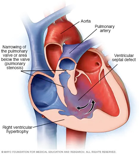
54. Tetralogy of Fallot (TOF) consists of four features: right ventricular outflow tract (RVOT) obstruction, a
large ventricular septal defect (VSD), an overriding ascending aorta, and RV hypertrophy.
55. The three Ds of the Ebstein anomaly are an apically displaced tricuspid valve that is dysplastic, with a
right ventricle that may be dysfunctional.
56. Systolic wall stress is described by the law of Laplace, which states that systolic wall stress is equal to:
(arterial pressure (p) × radius (r))/2 × thickness (h) , or σ = (p × r)/2h
57. Echocardiographic findings suggestive of severe mitral regurgitation include enlarged LA or LV, the
color Doppler mitral regurgitation jet occupying a large proportion (more than 40%) of the LA, a
regurgitant volume 60 mL or more, a regurgitant fraction 50% or greater, a regurgitant orifice
0.40 cm2 or greater, and a Doppler vena contracta width 0.7 cm or greater.
58. The seven factors that make up the Thrombolysis in Myocardial Infarction (TIMI) Risk Score are: age
greater than 65 years; three or more cardiac risk factors; prior catheterization demonstrating CAD;
ST-segment deviation; two or more anginal events within 24 hours; aspirin use within 7 days; and
elevated cardiac markers.
6 TOP 100 SECRETS
59. The components of the Global Registry of Acute Coronary Events (GRACE) Acute Cardiac Syndrome
(ACS) Risk Model (at the time of admission) are age; heart rate; systolic blood pressure, creatinine;
congestive heart failure (CHF) Killip class, ST-segment deviation; elevated cardiac enzymes and/or
markers; and presence or absence of cardiac arrest at admission.
60. Myocarditis is most commonly caused by a viral infection. Other causes include nonviral infections
(bacterial, fungal, protozoal, parasitic), cardiac toxins, hypersensitivity reactions, and systemic disease
(usually autoimmune). Giant cell myocarditis is an uncommon but often fulminant form of myocarditis
characterized by multinucleated giant cells and myocyte destruction.
61. Initial therapy for patients with non–ST segment elevation acute coronary syndrome (NSTEACS)
should include antiplatelet therapy with aspirin and with either clopidogrel, ticagrelor, or a
glycoprotein
IIb/IIIa inhibitor, and antithrombin therapy with either unfractionated heparin, enoxaparin,
fondaparinux, or bivalirudin (depending on the clinical scenario).
62. Important complications in heart transplant recipients include infection, rejection, vasculopathy (diffuse
coronary artery narrowing), arrhythmias, hypertension, renal impairment, malignancy (especially
skin cancer and lymphoproliferative disorders), and osteoporosis (caused by steroid use).
63. The classic symptoms of aortic stenosis are angina, syncope, and those of heart failure (dyspnea,
orthopnea, paroxysmal nocturnal dyspnea, edema, etc.). Once any of these symptoms occur, the
average survival without surgical intervention is 5, 3, or 2 years, respectively.
64. Class I indications for aortic valve replacement (AVR) include (1) development of symptoms in patients
with severe aortic stenosis; (2) an LV ejection fraction of less than 50% in the setting of severe aortic
stenosis; and (3) the presence of severe aortic stenosis in patients undergoing coronary artery bypass
grafting, other heart valve surgery, or thoracic aortic surgery.
65. The major risk factors for venous thromboembolism (VTE) include previous thromboembolism, immobility,
cancer and other causes of hypercoagulable state (protein C or S deficiency, factor V Leiden,
antithrombin deficiency), advanced age, major surgery, trauma, and acute medical illness.
66. The Wells Score in cases of suspected pulmonary embolism (PE) includes deep vein
thrombosis
(DVT) symptoms and signs (3 points); PE as likely as or more likely than alternative
diagnosis (3 points); heart rate greater 100 beats/min (1.5 points); immobilization or surgery in
previous 4 weeks (1.5 points); previous DVT or PE (1.5 points); hemoptysis (1.0 point); and cancer
(1 point).
67. The main symptoms of aortic regurgitation (AR) are dyspnea and fatigue. Occasionally patients
experience angina because reduced diastolic aortic pressure reduces coronary perfusion pressure,
impairing coronary blood flow. Reduced diastolic systemic pressure may also cause syncope or
presyncope.
68. The physical findings of AR include widened pulse pressure, a palpable dynamic LV apical beat that
is displaced downward and to the left, a diastolic blowing murmur heard best along the left sternal
border with the patient sitting upright and leaning forward, and a low-pitched diastolic rumble heard
to the LV apex (Austin Flint murmur).
69. Class I indications for aortic valve replacement in patients with AR include (1) the presence of symptoms
in patients with severe AR, irrespective of LV systolic function; (2) chronic severe AR with LV
systolic dysfunction (ejection fraction 50% or less), even if asymptomatic; and (3) chronic, severe AR
in patients undergoing coronary artery bypass grafting (CABG), other heart valve surgery, or thoracic
aortic surgery.
TOP 100 SECRETS 7
70. Cardiogenic shock is a state of end-organ hypoperfusion caused by cardiac failure characterized by
persistent hypotension with severe reduction in cardiac index (less than 1.8 L/min/m2) in the presence
of adequate or elevated filling pressure (LV end-diastolic pressure 18 mm Hg or higher or RV
end-diastolic pressure 10 to 15 mm Hg or higher).
71. The rate of ischemic stroke in patients with nonvalvular atrial fibrillation (AF) is about two to seven
times that of persons without AF, and the risk increases dramatically as patients age. Both paroxysmal
and chronic AF carry the same risk of thromboembolism.
72. In nuclear cardiology stress testing, a perfusion defect is an area of reduced radiotracer uptake in the
myocardium. If the perfusion defect occurs during stress and improves or normalizes during rest, it is
termed reversible and usually suggests the presence of inducible ischemia, whereas if the perfusion
defect occurs during both stress and rest, it is termed fixed and usually suggests the presence of scar
(infarct).
73. The main organ systems that need to be monitored with long-term amiodarone therapy are the lungs,
the liver, and the thyroid gland. A chest radiograph should be obtained every 6 to 12 months, and
liver function tests (LFTs) and thyroid function tests (thyroid-stimulating hormone [TSH] and free T4)
should be checked every 6 months.
74. The target international normalized ratio (INR) for warfarin therapy in most cases of cardiovascular
disease is 2.5, with a range of 2.0 to 3.0. In certain patients with mechanical heart valves (e.g., older
valves, mitral position), the target is 3.0 with a range of 2.5 to 3.5.
75. Lidocaine may cause a variety of central nervous system symptoms including seizures, visual disturbances,
tremors, coma, and confusion. Such symptoms are often referred to as lidocaine toxicity.
The risks of lidocaine toxicity are increased in elderly patients, those with depressed LV function, and
those with liver disease.
76. The most important side effect of the antiarrhythmic drug sotalol is QT-segment prolongation leading
to torsades de pointes.
77. The major complications of percutaneous coronary intervention (PCI) include periprocedural MI,
acute stent thrombosis, coronary artery perforation, contrast nephropathy, access site complications
(e.g., retroperitoneal bleed, pseudoaneurysm, arteriovenous fistula), stroke, and a very rare need for
emergency CABG.
78. The widely accepted hemodynamic definition of pulmonary arterial hypertension (PAH) is a mean
pulmonary arterial pressure of more than 25 mm Hg at rest or more than 30 mm Hg during exercise,
with a pulmonary capillary or LA pressure of less than 15 mm Hg.
79. Acute pericarditis is a syndrome of pericardial inflammation characterized by typical chest pain, a
pathognomonic pericardial friction rub, and specific electrocardiographic changes (PR depression,
diffuse ST-segment elevation).
80. Conditions associated with the highest cardiac risk in noncardiac surgery are unstable coronary
syndromes (unstable or severe angina), decompensated heart failure, severe valvular disease (particularly
severe aortic stenosis), and severe arrhythmias.
81. General criteria for surgical intervention in cases of thoracic aortic aneurysm are, for the ascending
thoracic aorta, aneurysmal diameter of 5.5 cm (5.0 cm in patients with Marfan syndrome), and
for the descending thoracic aorta, aneurismal diameter of 6.5 cm (6 cm in patients with Marfan
syndrome).
8 TOP 100 SECRETS
82. Cardiac complications of advanced AIDS in untreated patients include myocarditis and/or cardiomyopathy
(systolic and diastolic dysfunction), pericardial effusion/tamponade, marantic (thrombotic)
or infectious endocarditis, cardiac tumors (Kaposi sarcoma, lymphoma), and RV dysfunction from
pulmonary hypertension or opportunistic infections. Complications with modern antiretroviral therapy
(ART) include dyslipidemias, insulin resistance, lipodystrophy, atherosclerosis, and arrhythmias.
83. The radiation dose of a standard cardiac computed tomography (CT) angiography depends on a
multitude of factors and can range from 1 mSv to as high as 30 mSv. This compares to an average
radiation dose from a nuclear perfusion stress test of 6 to 25 mSv (or as high as more than 40 mSv
in thallium stress/rest tests) and an average dose from a simple diagnostic coronary angiogram of
approximately 5 mSv.
84. The ankle-brachial index (ABI) is the ankle systolic pressure (as determined by Doppler examination)
divided by the brachial systolic pressure. An abnormal index is less than 0.90. The sensitivity
is approximately 90% for diagnosis of peripheral arterial disease (PAD). An ABI of 0.41 to 0.90 is
interpreted as mild to moderate peripheral arterial disease; an ABI of 0.00 to 0.40 is interpreted as
severe PAD.
85. Approximately 90% of cases of renal artery stenosis are due to atherosclerosis. Fibromuscular
dysplasia (FMD) is the next most common cause.
86. In very general terms, in cases of carotid artery stenosis, indications for carotid endarterectomy
(CEA) are: (1) symptomatic stenosis 50% to 99% diameter if the risk of perioperative stroke or death
is less than 6%; and (2) asymptomatic stenosis greater than 60% to 80% diameter if the expected
perioperative stroke rate is less than 3%.
87. The most common cardiac complications of systemic lupus erythematosus (SLE) are pericarditis,
myocarditis, premature atherosclerosis, and Libman-Sacks endocarditis.
88. Cardiac magnetic resonance imaging (MRI) can be performed in most patients with implanted cardiovascular
devices, including most coronary and peripheral stents, prosthetic heart valves, embolization
coils, intravenous vena caval filters, cardiac closure devices, and aortic stent grafts. Pacemakers and
implantable cardioverter defibrillators are strong relative contraindications to MRI scanning, and scanning
of such patients should be done under specific delineated conditions, only at centers with expertise
in MRI safety and electrophysiology, and only when MRI imaging in particular is clearly indicated.
89. The clinical manifestations of symptomatic bradycardia include fatigue, lightheadedness, dizziness,
presyncope, syncope, manifestations of cerebral ischemia, dyspnea on exertion, decreased exercise
tolerance, and congestive heart failure.
90. Second-degree heart block is divided into two types: Mobitz type I (Wenckebach) exhibits progressive
prolongation of the PR interval before an atrial impulse (P wave) is not conducted, whereas Mobitz
type II exhibits no prolongation of the PR interval before an atrial impulse is not conducted.
91. Temporary or permanent pacing is indicated in the setting of acute MI, with or without symptoms, for
(1) complete third-degree block or advanced second-degree block that is associated with block in the
His-Purkinje system (wide complex ventricular rhythm) and (2) transient advanced (second-degree or
third-degree) AV block with a new bundle branch block.
92. Cardiac resynchronization therapy (CRT) refers to simultaneous pacing of both ventricles (biventricular,
or Bi-V, pacing). CRT is indicated in patients with advanced heart failure (usually NYHA class III
or IV), severe systolic dysfunction (LV ejection fraction 35% or less), and intraventricular conduction
delay (QRS less than 120 ms) who are in sinus rhythm and have been on optimal medical therapy.
TOP 100 SECRETS 9
93. Whereas the left internal mammary artery (LIMA), when anastomosed to the left anterior descending
artery (LAD), has a 90% patency at 10 years, for saphenous vein grafts (SVGs), early graft stenosis or
occlusion of up to 15% can occur by 1 year, with 10-year patency traditionally cited at only 50% to
60%.
94. Myocardial contusion is a common, reversible injury that is the consequence of a nonpenetrating
trauma to the myocardium. It is detected by elevations of specific cardiac enzymes with no evidence
of coronary occlusion and by reversible wall motion abnormalities detected by echocardiography.
95. Causes of restrictive cardiomyopathy include infiltrative diseases (amyloidosis, sarcoidosis, Gaucher
disease, Hurler disease), storage diseases (hemochromatosis, glycogen storage disease, Fabry
disease), and endomyocardial involvement from endomyocardial fibrosis, radiation, or anthracycline
treatment.
96. Classical signs for cardiac tamponade include the Beck triad of (1) hypotension caused by decreased
stroke volume, (2) jugulovenous distension caused by impaired venous return to the heart, and
(3) muffled heart sounds caused by fluid inside the pericardial sac, as well as pulsus paradoxus and
general signs of shock, such as tachycardia, tachypnea, and decreasing level of consciousness.
97. The most common tumors that spread to the heart are lung (bronchogenic) cancer, breast cancer,
melanoma, thyroid cancer, esophageal cancer, lymphoma, and leukemia.
98. Primary cardiac tumors are extremely rare, occurring in one autopsy series in less than 0.1% of
subjects. Benign primary tumors are more common than malignant primary tumors, occurring
approximately three times as often as malignant tumors.
99. The Westermark sign is the finding in pulmonary embolism of oligemia of the lung beyond the
occluded vessel. If pulmonary infarction results, a wedge-shaped infiltrate (Hampton’s hump) may be
visible.
100. Patients with cocaine-induced chest pain should be treated with intravenous benzodiazepines,
which can have beneficial hemodynamic effects and relieve chest pain, and aspirin therapy, as well
as nitrate therapy if the patient remains hypertensive. β-blockers (including labetalol) should not be
administered in the acute setting of cocaine-induced chest pain

Akathisia

Akathisia:

🔑In Greek means, “inability to sit”
🔑It is characterized by state of inner restlessness and shifting of weight. Hightened lower limb movements are quite characteristic
🔑It is an acute extrapyramidal symtom which typically is seen within 7-10 days of initiation of the antipsychotic
🔑Most common extrapyramidal side effect after starting an antpsychotic
🔑First generation older antipsychotics have higher chance of producing Akathisia compared to second generation antipsychotics
🔑Among the second generation antipsychotics, Clozapine has the least and Aripiprazole has the highest chance of causing Akathisia
🔑Management of Akathisia is:
💊If possible, the first step is to decrease the dose of antipsychotic
💊The commonly used drugs are Propranolol(most preferable), Benzodiazepines and Anticholinergics(least preferable)

Hypertensive treatment

When to initiate treatment for hypertension ??
JNC-8 RECOMMENDATIONS
---> For those more than or equal to 60 years, when SBP more than or equal to 150 mmHg or DBP more than or equal to 90 mmHg
--->For those with age less than 60 years, when SBP more than or equal to 140 mmHg or DBP more than or equal to 90 mmHg
---> For those with CKD and / or DM, when SBP more than or equal to 140 mmHg or DBP more than or equal to 90 mmHg (irrespective of age)

Sunday, 7 February 2016

ACUTE PANCREATITIS

RansonRanson’s Criteria on Admission :
age greater than 55 years
a white blood cell count of > 16,000/µL
blood glucose > 11 mmol/L (>200 mg/dL)
serum LDH > 350 IU/L
serum AST >250 IU/L

Ranson’s Criteria after 48 hours of admission :
fall in hematocrit by more than 10 percent
fluid sequestration of > 6 L
hypocalcemia (serum calcium < 2.0 mmol/L (<8.0 mg/dL))
hypoxemia (PO2 < 60 mmHg)
increase in BUN to >1.98 mmol/L (>5 mg/dL) after IV fluid hydration base deficit of >4 mmol/L

The prognostic implications of Ranson’s criteria are as follows :
Score 0 to 2 : 2% mortality
Score 3 to 4 : 15% mortality
Score 5 to 6 : 40% mortality
Score 7 to 8 : 100% mortality

APACHE
“Acute Physiology And Chronic Health Evaluation” (APACHE II) score > 8
points predicts 11% to 18% mortality
Hemorrhagic peritoneal fluid
Obesity
Indicators of organ failure
Hypotension (SBP <90 mmHG) or tachycardia > 130 beat/min
PO2 <60 mmHg
Oliguria (<50 mL/h) or increasing BUN and creatinine
Serum calcium < 1.90 mmol/L (<8.0 mg/dL) or serum albumin <33 g/L
(♥.2.g/dL)>

Balthazar Scoring for the Grading of Acute Pancreatitis
Grade A – normal CT
Grade B – focal or diffuse enlargement of the pancreas
Grade C – pancreatic gland abnormalities and peripancreatic inflammation
Grade D – fluid collection in a single location
Grade E – two or more collections and/or gas bubbles in or adjacent to pancreas

Monday, 11 January 2016

Endocrinology

Endocrine (Last Minute Revision)
1. diagnosis of diabetes mellitus Fasting BG>125 mg and post prandial >140mg, HbA1 >6.5%
2. Diabetes control is best monitored by: HbA1C
3. GLP-1 agonist, Exenatide
4. Insulin resistant if he needs more than – 200
5. Seen in DKA, Dehydration, Abdominal pain/tenderness
6. in DKA, Leukocytosis, Pseudohyponatremia occurs, Acute gastric dilatation
7. most dangerous complication of DKA, Cerebral edema
8. plasma of osmolality, 290
9. Deficiency of which of the following vitamins can lead to lactic acidosis , B1
10. The most reliable indicator for diabetic nephropathy is , Urine albumin >30 mg per day in 3 consecutive samples
11. Earliest feature of diabetic nephropathy, Raise GFR
12. Third nerve palsy with normal pupillary reaction, Diabetes
13. Drug used for DM gastropathy , Erythromycin
14. Drug is not used in erectile dysfunction, Apomorphine, Yohimbine, PgE1
15. Life threatening complications of diabetes mellitus are, Malignant otitis externa, Rhinocerebral mucormycosis, Emphysematous pyelonephritis
16. Weight loss may be seen in, Uremia, Pheochromocytoma , Adrenal insufficiency
17. parameter for Metabolic Syndrome (Syndrome X), Hypertriglyceridemia, Low HDL cholesterol, Hypertension
18. Hyperthyroidism following administration of supplemental iodine to subjects with endemic iodine deficiency goiter is known, Jod-Basedow effect
19. best marker to diagnose hypothyroid is, TSH
20. Decreased Radio iodine uptake is, Subacute thyroiditis , Factitious thyroiditis
21. ischemic heart disease [IHD] is diagnosed having hypothyroidism. Which of the following would be most appropriate line of management for, Start Levothyroxine at low dose
22. true about Hashimoto’s thyroiditis, Follicular destruction , Increase in lymphocytes, Oncocytic metaplasia , Orphan Annie eye nuclei
23. 'Hurthle cells' are seen, Hashimoto's Thyroiditis
24. feature of dequervan's disease, ESR, Painful & associated with enlargements of thyroid
25. common cause of hyperparathyroidism, Parathyroid adenoma
26. presentation of hyperparathyroidism is, Asymptomatic hypercalcemia
27. feature of hypercalcemia, Polyuria, Vomiting, Depression
28. Tufting of the terminal phalanges is seen in, Hyperparathyroidism
29. causes of hypercalcemia, Lithium therapy, Multiple myeloma, Vitamin A intoxication
30. true, Williams syndrome consists of precocious puberty, mental retardation & obesity
31. Hypercalcemia is seen in all, Sarcoidosis, Vitamin A toxicity, Phenytoin toxicity
32. Hypercalcemia is, Prolong immobilization,
33. Pseudohypoparathyroidism, ed PTH
34. Features of tumor lysis syndrome, Hyperuricemia, Hyperphosphatemia, Hyperkalemia
35. tumor lysis syndrome are, Hypocalcemia
36. Hypophosphatemia is seen in, Resolving phases of diabetic ketoacidosis, Respiratory alkalosis, Chronic alcoholism
37. Type I MEN involves, Pancreas, Pituitary, Parathyroid
38. associated with MEN 2, Pheochromocytoma, Medullary carcinoma thyroid, Parathyroid adenoma
39. features of MEN II b, Pheochromocytoma, Neuromas, Medullary ca thyroid
40. most common cause of Cushing's syndrome, Iatrogenic steroids
41. features of Cushing's disease, Central obesity, Easy bruising , Glucose intolerance
42. Cushing's disease is associated with, Increased ACTH and increased cortisol
43. earliest manifestation of Cushing's syndrome, Loss of diurnal variation
44. Conn's syndrome is most commonly associated with, Cortical adenoma
45. Conn's syndrome is associated with, Hypertension, Muscle weakness, Hypokalemia
46. cause pituitary apoplexy, Diabetes mellitus, Sickle cell anemia, Hypertension
47. Octreotide is useful for, Insulinoma, Glucagonoma, Carcinoid tumor
48. brain tumors spreads via CSF, Medulloblastoma
49. Heal pad thickness is increase in, Acromegaly
50. Commonest cause of increase heal pad thickness, Trauma
51. Hypoglycemia is seen in, Hepatoma
52. Hypercalcemia occur in, Breast, Bladder, Head & Neck
53. SIADH occur in, Small cell lung cancer
54. To diagnose a case of diabetes, the fasting blood sugar should be, 126
55. Type 1 diabetes is associated with, DR4
56. hyperglycemia occurs, Cushing syndrome
57. Hypoglycemia is a important feature, Addison disease
58. DPP IV inhibitor, Sitagliptin
59. Drug which can be used in both type I and type II diabetes, Acarbose
60. DKA case is treated with insulin the serum sodium level, Rise
61. Most common type of diabetic neuropathy, Sensory neuropathy
62. condition can lead to hypothyroid, Hemochromatosis
63. drug can lead to hypothyroid, Lithium
64. condition can lead hypothyroid, Scleroderma
65. Non pitting edema, Hypothyroid
66. Hung up reflex is seen, Hypothyroid
67. Which of the following is most common seen in hypothyroid, Raised LDL
68. Cyst in the bones are seen in, Hyperparathyroid
69. Edema feet is not a feature, Conn syndrome
70. Triad of pheochromocytoma includes, Palpitation, Headache, Sweating
71. Profuse sweating is a feature, Pheochromocytoma
72. Desmopressin is used in the treatment, Cranial diabetic insipidus
73. Hyponatremia occurs in which of, Small cell lung carcinoma

Tuesday, 29 December 2015

MODY

The most common MODY type is HNF1 alpha. This is responsible for 70% of MODY.

The amount of insulin produced by the pancreas becomes less as the person gets older, and MODY develops during adolescence or the early twenties.

Glucokinase is the second type of MODY, and occurs when this gene (that aids the body in recognising blood glucose levels) malfunctions.

This type of MODY can be hard to identify, and symptoms can be particularly slow in manifesting themselves.

It is usually picked up during routine testing. When a person is pregnant, it is important to screen for it.

HNF4 - alpha is a less common form of MODY that is often diagnosed at a later stage. HNF1 - beta is a type of MODY associated with renal cysts.

PDX1 and IPF1 are the same type of MODY, and are incredibly rare, affecting only one UK family to date.

NeuroD1 is another rare type of MODY, affecting only two families in the UK. Little information is available about the rarer forms of MODY.

Saturday, 26 December 2015

Wilsons Disease

Wilson's disease is an autosomal recessive disorder characterised by excessive copper deposition in the tissues. Metabolic abnormalities include increased copper absorption from the small intestine and decreased hepatic copper excretion. Wilson's disease is caused by a defect in the ATP7B gene located on chromosome 13.

Features result from excessive copper deposition in the tissues, especially the brain, liver and cornea:
• liver: hepatitis, cirrhosis
• neurological: basal ganglia degeneration, speech and behavioural problems are often the first manifestations
• Kayser-Fleischer rings
• renal tubular acidosis
• haemolysis
• blue nails

Diagnosis
• reduced serum caeruloplasmin
• increased 24hr urinary copper excretion

Tuesday, 22 December 2015

IgA Nephropathy

IMPORTANT FACTS –
IgA nephropathy is the most common form of glomerulonephritis worldwide.
There is male preponderance, peak incidence is in the second & third decades of life.
Two most common clinical presentations are:
● Recurrent episodes of macroscopic hematuria.
● Persistent asymptomatic microscopic hematuria.
Renal histological features of HSP nephritis is strikingly similar to those of IgA nephropathy.


IgA Nephropathy

IMPORTANT POINTS
Aphemia is a severe form of acute speech apraxia that presents with severely impaired fluency.
‪Warfarin is not contraindicated in lactating mothers.
Serum lithium level should be checked earliest after 5 days of constant dosing.
Gamma Gandy bodies contain hemosiderin & calcium.
Internucleosomal cleavage of DNA is characteristic of apoptosis.
Most common form of dissociation hysteria is amnesia.
Relapse rate of P.vivax malaria in India is 30%.
Radiation dose of barium enema is equal to 350 X-rays.
Ram's horn & Pseudobillroth appearance on barium studies is a feature of Crohn's disease.
Thiazides decrease positive free water clearance.
Carbohydrate, protein & fat metabolism occur in TCA cycle.
Monosaccharides seperation is by chromatography technique.
Papillary hidradenoma occurs most commonly in anogenital region.
Most common location for performing endomyocardial biopsy is right ventricle.
Earliest immunoglobulin to appear in fetus is IgM.
Cow milk contains soluble radioactive substance - Strontium 90.
Earliest visual field defect in Glaucoma is isopter contraction but paracentral scotomas are more specific.
BASIC FACTS
Paracrine regulation refers to factors released by one cell that act on an adjacent cell in the same tissue.
For example, somatostatin secretion by pancreatic islet delta cells inhibits insulin secretion from nearby beta cells.
Autocrine regulation describes the action of a factor on the same cell from which it is produced. IGF-1 acts on many cells that produce it, including chondrocytes, breast epithelium & gonadal cells.

Thursday, 15 October 2015

DKA

DIABETIC KETO ACIDOSIS is life threatening complication in diabetic patients who need insulin therapy. I stress again who needs insulin therapy.....
KETOACIDOSIS occurs more often in type I diabetic patients no matter how good is the compliance with the insulin therapy. One precipitating factor and the DKA happens. Contrary to this patients with type II Diabetes Mellitus develop DKA only either when they have insulin secreating islet beta cells exhausted so on insulin therapy or when the precipitation is either multifactorial or very intense.
The pathophysiology behind is the fact that although the total insulin released in type II DM is much less compared to type I DM but whatever insulin is released is in the portal system and directly reaches the site of ketogenesis i.e liver and thus prevents ketogenesis in the mitochondria of the hepatocytes while in type I most of the insulin injected in periphery hardly reach the portal system while being utilised in the periphery especially in precipitating situations for DKA like Ischemia(Cardiovascular, Cerebrovascular and peripheral vascular), infarction, inadequate insulin , pregnancy and drugs like cocain

Saturday, 19 September 2015

ASD

Atrial Septal Defect (ASD)

MC- Secundum (Center)✅
Primum (big on medial side)✅
Sinus Venosus (at insertion of SVC, IVC)✅

Seen in
Down's Syndrome✅
Lutembacher's syndrome ( ASD + MS/MR)✅
Ellis van creveld syndrome ( ASD + Polydactyly)✅
Holt oram syndrome ( Autosomal Dominant )✅

🍀Holt Oram Syndrome = Hand heart syndrome :

🌱Familial ASD syndrome

🌱With - VSD , Primary heart block , Atrial fibrillation, Bony Abnormality ( Thumb - absent / rudimentary)

🌱Pleiotropy - Multiple effects due to defect in single gene ( here TBX5)
🌱Can have absent radius.

🍀ASD Secundum :
1. Asymptomatic
2. S2 - Wide and fixed split
3. Grows up -
Adult -
CHF
Arrhythmias
Stroke
Eisenmenger syndrome

🌱ECG : Right axis deviation.

Treatment : Closure at 3-5yrs age by Dacron Patch

🍀ASD Primum :

1. Big cleft on the medial side
2. Also has MR (always)
Pansystolic at Apex
Radiate to axila and back
3. S2 - Wide and fixed split
4. It is severe ASD

therefore

Clinical features at 6 - 10 wks
Failure to thrive
Recurrent infections
Feeding difficulties
CHF
ECG : Left axis deviation.

Treatment : Early surgery.

Tuesday, 7 July 2015

Mutations in MODY

Mutations in MODY
Type 1: HNF 4 alpha
Type 2: glucokinase
Type 3: HNF 1 alpha
Type 4: IPF1 (insulin promoter factor 1)
Type 5: HNF 1 beta