Friday, 22 June 2018

Signs in ENT


BATTLE SIGN- Bruising behind earat mastoid region, due to petroustemporal bone# (middle fossa #).

BOCCA'S SIGN - Absence of postcricoid crackle(Muir's crackle) inCarcinoma post. cricoid.

BROWN SIGN - blanching of rednesson increasing pressure more thansystemic pressure see in glomusjugulare.

BOYCE SIGN - Laryngocoele-Gurgling sound on compression ofexternal laryngocoele with reductionof swelling.

DODD’S SIGN/CRESCENT SIGN - X-ray finding-Crescent of air betweenthe mass and posterior pharyngealwall. positive in AC ployp. Negativein Angiofibroma

FURSTENBERGERSSIGN-This is seenwhen nasopharyngeal cyst is communicating intracranially,there isenlargement of the cyst on crying and upon compression of jugularvein.

HITSELBERGER'SSIGN - In Acousticneuroma- loss of sensation in theear canal suppllied by Arnold'snerve( branch of Vagus nerve to ear )

HOLMAN MILLER SIGN, ANTRALSIGN-it is seen in angiofibroma,thetumor pushes forward on theposterior wall of the maxillarysinus..

HONDOUSA SIGN--X-ray finding inAngiofibroma, indicatinginfratemporal fossa involvementcharacterised by widening of gapbetween ramus of mandible andmaxillary body.

HENNEBERT SIGN- false fistula sign( cong.syphilis, Meniere's,)

IRWIN MOORE’S SIGN-------- positivesqueeze test in chronic tonsillitis

LIGHT HOUSE SIGN--- seeping outof secretions in acute OTITIS media

LYRE'S SIGN - splaying of carotidvessels in carotid body tumor

MILIAN’S EAR SIGN- Erysipelas canspread to pinna(cuticularaffection),where as cellulitis cannot.

PHELP'S SIGN - loss of crust of bonebetween carotid canal and jugularcanal in glomus jugulare

RACOON SIGN-Indicate subgalealhemorrhage,and not necessarly baseof skull #

STEEPLE SIGN- X-ray finding inAcute Laryngo tracheo bronchitis

STANKIEWICK'S SIGN - indicateorbital injury during FESS. fatprotrudes into nasal cavity oncompression of eye ball from ouside

THUMB SIGN --X-ray finding A/cepiglottitis

TRAGUS SIGN- EXTERNAL OTITIS ,Pain on pressing Tragus

TEA POT SIGN is seen in CSFrhinorrhoea..

WOODS SIGN----- palpable jugulodigastric lymphnode

Tuesday, 12 June 2018

Tetralogy of Fallot, demystified


Tetralogy of Fallot is a fairly common heart defect. In fact, it's the most common cyanotic heart defect (meaning that if a baby is born with cyanosis - markedly decreased oxygen saturation - and the cause is determined to be a congenital heart defect, the most likely culprit is tetralogy of Fallot).

From the name, it’s obviously composed of four parts. But how to remember what those parts are? You could just memorize them using brute force, but there's actually one thing that ties them all together - so if you can remember this one thing, then the four things make sense. I love this, because when you find you've forgotten one of the four things (which you probably will), you can actually reason it out. Hurray!

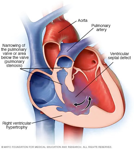
Here's the one thing to remember, and it's actually the thing that causes the whole disease. When the interventricular septum is forming, the top portion is pushed up and towards the right ventricle. The official name for this is anterosuperior displacement of the infundibular septum, but it's easier to just remember "up and towards the right," I think.


Check out this image of tetralogy of Fallot. See the red asterisk? That's the top of the interventricular septum. The black asterisk marks the bottom of the interventricular septum. Those parts are supposed to be connected...but obviously they're not. And the reason they're not is that the top part of the septum has moved up (creating a hole in the septum) and to the right (smushing the pulmonary artery outflow tract).

So what happens as a result?

One of the main problems is that it's now very hard for the blood to get out of the right ventricle and into the pulmonary artery (and lungs). That's because the pulmonary outflow tract (fancy name for the beginning of the pulmonary artery) is compressed, and there's less room for blood to flow through. This is called pulmonary stenosis. This is what causes the baby to be cyanotic! If you can't get enough blood out into the lungs, the blood isn't going to be oxygenated very well, and the baby's skin will be bluish. The right ventricle has to work hard to push blood through that compressed pulmonary artery - so the right heart becomes hypertrophied (bigger).

A couple other things happen too, as a result of this displacement of the septum. As the top part of the septum moves upwards, it separates from the bottom part, creating a hole in the septum (this is called a ventricular septal defect). This is actually kind of a good thing, in this case, because it relieves a little of the pressure on the right side of the heart. If the septum was intact, then the only place for the right ventricular blood to go would be through the compressed pulmonary artery...and the right ventricle would have to work incredibly hard to empty itself with each cardiac cycle.

Finally, as the top part of the septum moves to the right, it pulls the aortic valve along with it, repositioning it so that it sits pretty much right over the ventricular septal defect. This is called an overriding aorta, and it doesn't have much clinical consequence.  

So to summarize: the top of the septum moves up and to the right, causing:
1. Pulmonary stenosis 
2. Right ventricular hypertrophy 
3. A ventricular septal defect
4. An over-riding aorta  

Sunday, 27 May 2018

Fistula test Interpretation

How are the results of Fistula test interpreted?
1. In fistula over the dome of the lateral semicircular canal: Increase pressure causes conjugate horizontal deviation of the eyes towards the normal side. As pressure is maintained, jerk nystagmus develops with fast component towards the affected ear. As pressure is released, eyes return to normal
2. Fistula of the lateral semicircular canal (anterior to the ampulla) causes deviation of eyes, to the affected side
3. Vestibular erosion causes rotatory horizontal nystagmus towards the diseased ear
4. Fistula of the posterior semicircular canal causes vertical movement of eyes.

https://youtu.be/4gEM17yTl5k

Tuesday, 22 May 2018

Drowning

Drowning
Drowning is a major cause in head injuries and death
• Initial peak
– Toddler age group
• Second peak
– Male adolescents
• Children younger than 1 year of age
– Often drown in bathtubs, buckets, and toilets
• Children 1–4 years of age
– Likely drown in swimming pools where they haveb beenunsupervised temporarily (usually for < 5 min)
– Typical incidents involve a toddler left unattendedt temporarilyor under the supervision of an older sibling
• Adolescent and young adult age groups (ages 15–24 years)
– Most incidents occur in natural water
• Approximately 90 % of drowning occur within 10 yardso ofsafety
• Parent should be within an arm’s length of a swimmingc hild (anticipatory guidance)
Mechanism of injury
• Initial swallowing of water
• Laryngospasm
• Loss of consciousness
• Hypoxia
• Loss of circulation
• Ischemia
• CNS injury (the most common cause of death)
• Acute respiratory distress syndrome (ARDS) may develop
• Salt water drowning classically associated with:
– Hypernatremia
– Hemoconcentration
– Fluid shifts and electrolyte disturbances are rarely seenc linically
• Fresh water drowning classically associated with:
– Hyponatremia and hemodilution
– Hyperkalemia
– Hemoglobinuria and renal tubular damage
• Management of drowning and near drowning
– Cardiopulmonary resuscitation (CPR) at the scene
– Admit regardless of clinical status
– All children with submersion should be monitoredi inthe hospital for 6–8 h
– If no symptoms develop can be discharged safely
– 100 % oxygen with bag and mask immediately
– Nasogastric tube for gastric decompression
– Cervical spine immobilization if suspected cervicali injuries
– Positive end expiratory pressure (PEEP) and positivep ressure ventilations in case of respiratory arrest
– Continuous cardiac monitoring
– Bolus of normal saline or Ringer’s lactate
– Vasopressors
– Defibrillation if indicated

Monday, 12 February 2018

Mnemonics in Dermatology

Generalized Skin Hyperpigmentation

"With generalized, none of the skin is SPARED"

Sunlight

Pregnancy

Addison's disease

Renal failure

Excess iron (haemochromatosis)

Drugs (e.g. amiodarone, minocycline)

Painful Cutaneous Nodules

BENGAL CO.

Blue rubber bleb nevus

Eccrine spiradenoma

Neurilemmoma/Neuroma

Glomus tumor

Angiolipoma/Angioleiomyoma/Angiosarcoma

Leiomyoma

Cutaneous endometriosis/Calcinosis cutis

Osteoma cutis

White Patch of Skin

"Vitiligo PATCH"

Vitiligo

Pityriasis alba/Post-inflammatory hypopigmentation

Age related hypopigmentation (e.g. idiopathic guttate hypomelanosis)

Tinea versicolor, Tuberous sclerosis (ash-leaf macules)

Congenital birthmark (e.g. Hypomelanosis of Ito)

Hansen's disease (leprosy)

Common Causes of Leukocytoclastic Vasculitis

VASCULITIS

Viral (e.g. Hepatitis B and C)

Autoimmune (Systemic Lupus Erythematosus, Sjögren's syndrome, rheumatoid arthritis)

Streptococci, Staphylococci, Henoch-Schönlein purpura

Cryoglobulins, Cryofibrinogens, Churg Strauss/Wegener's granulomatosis

Ulcerative colitis, urticarial vasculitis

Lymphoproliferative disease (hairy cell leukemia)

Infectious (endocarditis, meningococcemia, gonococcemia, Rocky Mountain spotted fever)

Thiazides, phenothiazines, and other drugs

Immune complex reactions, iodides, idiopathic

Sulfa drugs (septra), penicillin, and other antibiotics

Non-scarring alopecia

TOP HAT

Telogen effluvium, Tinea capitis

Out of iron, zinc

Physical-trichotillomania, traction alopecia

Hormonal-hypothyroidism, androgenic

Autoimmune-alopecia areata, anagen effluvium

Toxins-heavy metals, chemotherapy

Erythema Nodosum

NODOSUM5

NO cause is found in 60% of cases

Drug (iodides, bromides, sulfonamides)

Oral contraceptives

Sarcoidosis or. Löfgren's syndrome

Ulcerative colitis, Crohn's disease, Behçet's

Microbiology: any chronic infection (bacterial, viral, yersinia, tuberculosis, leprosy, deep fungal)

Behcet's syndrome: Diagnostic Criteria

PROSE

Pathergy test

Recurrent genital ulceration

Oral ulceration (recurrent)

Skin lesions (e.g. erythema nodosum, subcutaneous throm-bophlebitis, cutaneous hypersensitivity)

Eye lesions (e.g. iridocyclitis, chorioretinitis)

• Oral ulceration is central criterion, plus any 2 others

Focal Dermal Hypoplasia Syndrome/Goltz syndrome

FOCAL

Female sex (85-90%)6

Osteopathia striata

Coloboma

Absent ecto-, meso-, and neuro-dermis elements

Lobster claw deformity

SLE (Systemic Lupus Erythematosus) Diagnosis

SOAP BRAIN MD

Serositis

Oral/nasal ulcers

Arthritis

Photosensitivity

Blood (cytopenia)

Renal involvement

ANA

Immune (typical antibodies e.g. dsDNA, anti-Sm)

Neurologic (e.g. seizures, stroke)

Malar rash

Discoid rash

• 4 out of 11 criteria needed for diagnosis

Henoch-Schonlein purpura: signs and symptoms

NAPA

Nephritis

Arthritis, arthralgias

Purpura , palpable (especially on lower extremities)

Abdominal pain (intussusception to be ruled out)

Monday, 15 January 2018

Anatomy One Liners

Anatomy One-Liners
1) Ascending colon 12.5cm long
- from the caecum to the inferior surface of  the right lobe of the liver
- usually retroperitoneal
(2) Transverse colon 50cm long 
- from the right colic flexure to the left colic flexure
- suspended by transverse mesocolon attached to the anterior border of the pancreas
(3) Descending colon - 25cm long
- from left colic flexure to the sigmoid colon
- it is narrower than ascending colon
- usually, it is retroperitoneal
(4) Sigmoid colon 37.5cm long
- from pelvic brim to the third piece of the sacrum, where it becomes rectum
- suspended by sigmoid mesocolon


Mesentery:- The mesentery of the small intestine (or) mesentery proper is a broad, fan-shaped fold of peritoneum which suspends the coils of jejunum and ileum from the posterior abdominal wall
Root of mesentery - 15cm long
- directed obliquely downwards and to the right
- It extends from the duodenojejunal flexure on the left side of vertebra L2 to the upper part of the right sacroiliac joint
- It crosses the following :
(1) Third part of duodenum where the superior mesenteric vessels enter into it
(2) The abdominal aorta
(3) The inferior vena cava
(4) The right ureter
(5) The right psoas major

The free or intestinal border is 6meter long, thrown into pleats.


Development of diaphragm by
1. Septum transverum
2.pleuro peritoneal membrane
3.somites (3-5th)
4.ventral pleural sac
5.mesentry of esophagus

Tonsil develop embryologically from 2nd pharyngeal pouch
Parathyroid is developed from 3rd & 4th brachial pouches
Footplate of stapes is developed from otic capsule
Umbilical vesicle attain full development in 4th week of fetus
Urachal fistula result from persistent allantois
Tensor tympani and tensor palatine supplied by trigeminal.Levator palatine supplied by the superior laryngeal nerve.
Derivatives of pharyngeal pouch
1st- tubotympanic recess
2nd-tonsil
3rd-inf parathyroid, thymus
4th-sup parathyroid, thyroid
5th-ultimobranchial body

Mesodermal derivatives of branchial arch
1st-malleus, incus, anterior ligament of malleus, sphenomandibular ligament
2nd-stape, styloid process, stylohyoid ligament, lesser cornu of hyoid, sup part of the hyoid
3rd-greater cornu of hyoid, lower part of the body of hyoid
4n6th-laryngeal cartilage
1st pharyngeal arch cartilage-Meckel cartilage
2nd pharyngeal arch cartilage-Reichert cartilage

TYMPANIC membrane dev from all the three germ layers
Pinna dev from 1st n 2nd pharyngeal arches
Footplate and annular ligament from otic capsule
Left umbilical vein-ligamentum teres
Rt-disappear
Ductus afteriosus-ligamentum arteriosum
Ductus venosus-ligamentum venosum
Septum primum n secondum-fossa ovalis n annulus ovalis respective
Distal umbilical art-median umb ligament
Proximal -sup vesical art
Foregut forms -Oesophagus
- The stomach
- Upper part of duodup to upto the opening of
common bile duct
Midgut forms - Rest of the duodenum
- Jejunum
- The ileum
- The appendix
- The caecum
- The ascending colon
- The right two - thirds of transverse colon
Hindgut forms -Left one-third of transverse colon
- The descending colon
- The sigmoid colon
- Proximal upper part of the rectum

Nerve supply of anal canal
(1) Above the pectinate line
- Sympathetic - Inferior hypogastric plexus - L1 & L2
- Parasympathetic- pelvic splanchnic S2, S3, S4
- Pain is carried by both of them
(2) Below the pectinate line
- somatic
- Inferior rectal S2,S3,S4 - nerves
(3) Sphincters - Internal sphincter - contraction - sympathetic nerve / relaxation - parasympathetic. N
External sphincter - inferior rectal nerve & by perineal branch of 4th sacral. N

Genital tubercle forms - clitoris
- urethral folds forms - labia minora
- genital swelling form - labia majora
- urogenital membrane gets ruptured to form the vestibule

Tuesday, 29 August 2017

Forebrain developmental abnormalities

Megalencephaly
  • Abnormally large brain
  • Less common than microencephaly 
Microencephaly
  • Abnormally small brain
  • Lots of causes (chromosomal abnormalities, fetal alcohol syndrome, HIV acquired in utero)
Lissencephaly
  • Decrease in number of gyri
  • Sometimes gyri are totally absent (agyria)
Polymicrogyria
  • Small, numerous, irregularly formed gyri
  • Can be caused by injury or genetic abnormality
Neuronal heterotopias
  • Neurons in inappropriate locations along migrational pathways
  • Associated with epilepsy
Holoprosencephaly
  • Incomplete separation of cerebral hemispheres across the midline
  • May have midline facial abnormalities (e.g., cyclopia, absence of olfactory cranial nerves)
Agenesis of the corpus callosum

  • Absence of white matter bundles connecting cerebral hemispheres
  • Patients may have other defects, or may be asymptomatic